Tissue detection device in minimally invasive surgery

A detection device and minimally invasive surgery technology, applied in the field of medical devices, can solve the problems that affect the wide application of liquid biopsy technology and high detection cost, and achieve the effect of eliminating hidden dangers, reducing pain, and lowering requirements

Pending Publication Date: 2020-07-17
胡三元
View PDF4 Cites 0 Cited by
  • Summary
  • Abstract
  • Description
  • Claims
  • Application Information

AI Technical Summary

Problems solved by technology

However, the existing liquid biopsy technology has a low number and concentration of circulating tumor cells (CTC) or circulating tumor DNA (ctDNA) in the blood of patients in the early stage of tumors, and is often lower than the detection limit, so that liquid biopsy technology is not effective in early tumors. In addition, liquid biopsy technology mainly relies on the separation of cancer cells in blood, and its detection cost is high. For patients who need continuous detection to achieve follow-up treatment, the medical burden of high cost will increase Daunting for patients and their families, but also affecting the widespread use of liquid biopsy technology

Method used


Image

  • Tissue detection device in minimally invasive surgery
  • Tissue detection device in minimally invasive surgery
  • Tissue detection device in minimally invasive surgery

Examples


Embodiment 1

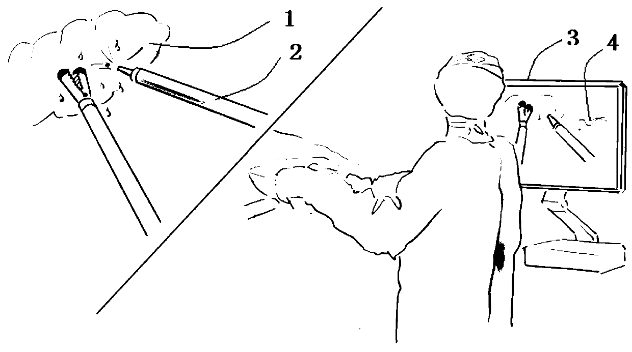
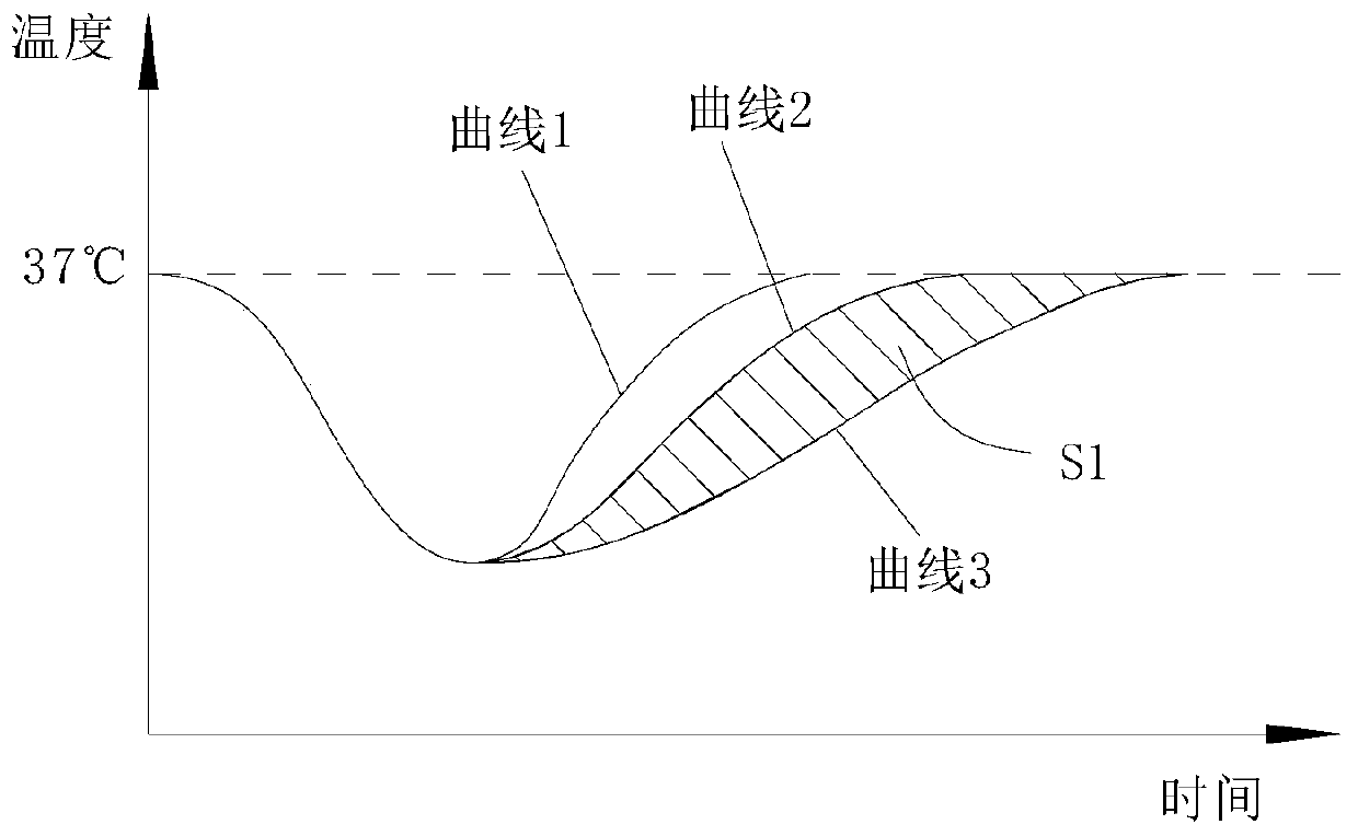
[0049] Embodiment 1, the feature quantity is an image used to characterize the temperature change over time of the tissue to be inspected 1 after it is sprayed with a cooling medium, for example, as figure 1 and figure 2 As shown in , it gives the characteristic quantity as a temperature-time variation curve used to characterize the temperature rise rate of the tissue 1 to be tested. Wherein, the horizontal axis is time, and the vertical axis is temperature. Curve 1 is a graph of the temperature of abnormal tissue changing with time, and curves 2 and 3 are graphs of the temperature of normal tissue changing with time.

Embodiment 2

[0050] In embodiment 2, the characteristic quantity is the time it takes for the temperature of the tissue 1 to be tested to rise to a fixed temperature after being sprayed by the cooling medium. This embodiment does not specifically limit the fixed temperature. As a preference, the fixed temperature is 36-37°C.

Embodiment 3

[0051] In embodiment 3, the characteristic quantity is the temperature of the tissue to be tested 1 detected at a certain time point (eg, 15 minutes) after the tissue to be tested is sprayed with a cooling medium.


PUM


Abstract

The invention discloses a tissue detection device in minimally invasive surgery. The tissue detection device comprises an insertion unit and a display unit; the insertion unit comprises an insertion tube and an integrated section connected with the insertion tube; the insertion unit also comprises a cooling medium spraying component and an endoscope component; the cooling medium spraying componentcomprises a spray head for spraying a cooling medium into an operation object body and a catheter communicating with the spray head; the endoscope component comprises a camera element and a temperature detection element; the camera element is used for acquiring tissue images; the temperature detection element is used for detecting the temperature of tissues in the operation object body; the sprayhead, the camera element and the temperature detection element are integrated in the integrated section; and the display unit are used for displaying the tissue images and the characteristic quantitycapable of representing the temperature rise rate of the tissues to be detected. According to the detection device disclosed by the invention, tissues in an operation can be rapidly detected throughthe temperature change rate under an operation condition, so that identification of tumor tissues is realized, and the detection device has the advantages of being minimally invasive, being capable ofrealizing real-time dynamic detection and continuous detection, and the like.

Description

technical field [0001] The present application relates to the technical field of medical devices, in particular to a tissue detection device during minimally invasive surgery. Background technique [0002] Tumor is an abnormal lesion formed by the body under the action of various carcinogenic factors when a certain cell in a local tissue loses its normal regulation of its growth at the gene level, resulting in its clonal abnormal proliferation. At present, there are various types of tumor detection methods, such as laboratory examination, radiological examination, radionuclide examination, ultrasonic examination, etc. There is one commonality in the existing various tumor detection methods, that is, pathological analysis of tumors requires sampling The two paths of analysis and image analysis have defects such as long inspection cycle, inability to make a final diagnosis, and follow-up observation. In particular, the sampling and analysis process for the purpose of confirmi...

Claims


Application Information

Patent Timeline
IPC IPC(8): A61B5/01A61B1/04A61B1/12A61B1/07
CPCA61B5/01A61B1/04A61B1/128A61B1/07
Inventor 胡三元
Owner 胡三元
Why Eureka
Try Eureka