Multi-color fluorescent probe based on DNA nanostructure as well as preparation method and application of multi-color fluorescent probe

A technology of fluorescent probes and nanostructures, which is applied in the field of multicolor fluorescent probes based on DNA nanostructures and its preparation, can solve the problems of luminescent properties (uncontrollable color and intensity, low number of fluorescent probe codes, etc.), and achieve good results. Biomedical application prospects, the effect of good biocompatibility

Active Publication Date: 2020-07-24
SHANGHAI ADVANCED RES INST CHINESE ACADEMY OF SCI
View PDF5 Cites 8 Cited by
  • Summary
  • Abstract
  • Description
  • Claims
  • Application Information

AI Technical Summary

Problems solved by technology

[0004] The purpose of the present invention is to provide a multicolor fluorescent probe based on DNA nanostructure and its preparation method and application, so as to sol...

Method used


Image

  • Multi-color fluorescent probe based on DNA nanostructure as well as preparation method and application of multi-color fluorescent probe
  • Multi-color fluorescent probe based on DNA nanostructure as well as preparation method and application of multi-color fluorescent probe
  • Multi-color fluorescent probe based on DNA nanostructure as well as preparation method and application of multi-color fluorescent probe

Examples


Embodiment 1

[0045] The present invention provides a multicolor fluorescent probe based on DNA nanostructure and its preparation method and application. Firstly, the preparation of DNA tetrahedron includes the following steps:

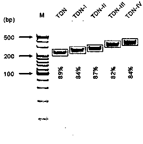
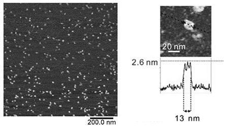
[0046] Dissolve all DNA in Q water, measure the absorption of single strands at 260 nm by an ultraviolet spectrophotometer, and quantify the concentration of all single strands of DNA to 100 μM. Mix equal amounts of four single-stranded DNAs for preparing DNA tetrahedrons in TM buffer (10 mM Tris-HCl, 5 mM MgCl 2 , PH 8.0), the final concentration of single-stranded DNA is 1 μM. Keep the temperature at 95 ℃ for 10 min in the PCR machine, then quickly lower the temperature to 4 ℃, and keep it at 4 ℃ for 10 min to prepare the DNA tetrahedron. Then, 8% polyacrylamide electrophoresis and AFM were used to characterize the DNA tetrahedrons. The electrophoresis running conditions were 120 V for 120 min. The morphology of DNA tetrahedron was characterized by AFM.

[0047] Res...

Embodiment 2

[0049] The preparation of high-order DNA tetrahedral self-assembly structure includes the following steps:

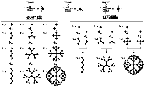
[0050] The DNA tetrahedrons modified with complementary arm chains were mixed according to the assembly molar ratio of 1:1, and incubated at 37°C for 2h to obtain the first-generation high-order DNA tetrahedral self-assembly structure (G1). Then, the second generation (G2), third generation (G3), and fourth generation (G4) high-order DNA tetrahedral self-assembly structures were synthesized by incubating at 37°C for 2 h by layer-by-layer assembly or fractal assembly. The assembly rules of high-order DNA tetrahedrons are as follows. From the central tetrahedron a-T0 as the assembly center, each additional layer represents the generation of a new generation of high-order structures. The n-1th generation tetrahedral high-order structure a-Gn-1 with arm chains is 3×2 (n-1), and the nth generation is obtained by hybridization with tetrahedrons with complementary hybrid arm chai...

Embodiment 3

[0059] Using the structural characteristics of the high-order DNA tetrahedral self-assembly structure, we further studied whether it is possible to construct multicolor fluorescent probes without crosstalk between adjacent molecules and quantifiable fluorescence intensity through the labeling of fluorescent molecules. Each DNA tetrahedron in the high-order DNA tetrahedron self-assembly structure has a site for labeling fluorescent molecules, so each tetrahedron can label 4 fluorescent molecules. We choose Alexa488, ROX and Cy5 three kinds of fluorescent molecules to construct multicolor fluorescent probes. For the F3,2 structure, 28 fluorescent molecules can be labeled.

[0060] Fluorescence spectra of the constructed fluorescent probes were collected using Edinburgh FS920 fluorescence spectrophotometer. The concentration of all tested samples was 10 nM. Alexa 488 excitation wavelength is 488 nm, spectral reception range is 495-590 nm; ROX excitation wavelength is 588 nm, spect...


PUM


Abstract

The invention discloses a multi-color fluorescent probe based on a DNA nanostructure as well as a preparation method and application thereof. The method comprises the following steps: constructing DNAtetrahedrons with different arm chain numbers; constructing a DNA nano self-assembly structure by a strategy of layer-by-layer assembly or fractal assembly, and marking fluorescent molecules of different types and quantities on the DNA nano self-assembly structure so as to construct the multicolor fluorescent probe based on the DNA nano self-assembly structure. According to the invention, the small-size multicolor fluorescent probe is constructed by a self-assembly means from bottom to top, and the defects of low coding quantity and uncontrollable optical property of the traditional fluorescent label are overcome. The multi-color fluorescent probe provided by the invention can be used for monomolecular level target molecule recognition and tumor cell recognition, and has good biocompatibility, so that the multicolor fluorescent probe has a good biomedical application prospect.

Description

Technical field [0001] The invention belongs to the field of biotechnology, and specifically relates to a multicolor fluorescent probe based on a DNA nanostructure, and a preparation method and application thereof. Background technique [0002] The simultaneous qualitative or quantitative detection of a variety of small biological molecules, nucleic acids, and proteins has important scientific significance for basic research in life sciences and biomedicine. For example, the occurrence of complex diseases such as malignant tumors involves a variety of biochemical reactions in the body, and diseases usually have multiple associated biomarkers. Therefore, compared to the detection of a single marker, multi-channel detection and imaging of multiple markers can improve the accuracy of early detection results and reduce false positive or false negative detection results. Therefore, multi-channel detection is an important method for the study of physiological and pathological processe...

Claims


Application Information

Patent Timeline
IPC IPC(8): C09K11/06C07H21/04G01N21/64
CPCC09K11/06C07H21/04G01N21/6486C09K2200/0476
Inventor 王丽华李江代江兵谢茉翟婷婷樊春海
Owner SHANGHAI ADVANCED RES INST CHINESE ACADEMY OF SCI
Why Eureka
Try Eureka