Bedside pulmonary ventilation-blood perfusion impedance tomography method based on saline radiography

A technology of electrical impedance tomography and imaging method, applied in the field of clinical medicine, can solve the problems of affecting imaging analysis, poor applicability, time-consuming, etc., and achieve the effect of improving efficiency, reducing interference, and high clinical application value

Active Publication Date: 2020-07-28
PEKING UNION MEDICAL COLLEGE HOSPITAL CHINESE ACAD OF MEDICAL SCI
View PDF7 Cites 7 Cited by
  • Summary
  • Abstract
  • Description
  • Claims
  • Application Information

AI Technical Summary

Problems solved by technology

[0004] At present, the quality of bedside pulmonary vascular perfusion imaging based on electrical impedance technology (EIT) is poor, and it is interfered by electrical resistance signals such as heart beating and pulmonary vascular pulsation, so its clinical application is poor.
In order to improve the imaging quality, some studies have developed the electrical impedance imaging method of saline contrast technology. Saline is injected through the central venous catheter, and at the same time apnea, the change of thoracic resistance signal is collected, and the local resistance-time change curve of saline contrast is established, but the resistance- There are many types of time curve analysis methods. During the saline injection process, it is difficult to identify the arrival of saline into the right atrium, reduce the interference of the resistance signal, and affect the imaging analysis. It is difficult to obtain the accurate time point of the first arrival of saline in the lung. , to determine the inconsistency between the time when the lung resistance drops and the heart resistance drops in a certain area, but the practicability is poor and time-consuming
[0005] Bedside pulmonary blood perfusion imaging analysis, the current electrical impedance technology mostly focuses on the distribution percentage of ventilation and blood flow, which belongs to semi-quantitative evaluation, and it is technically difficult to go deep into the equivalent quantitative calculation of ventilation dead space and intrapulmonary shunt

Method used


Image

  • Bedside pulmonary ventilation-blood perfusion impedance tomography method based on saline radiography
  • Bedside pulmonary ventilation-blood perfusion impedance tomography method based on saline radiography
  • Bedside pulmonary ventilation-blood perfusion impedance tomography method based on saline radiography

Examples


Embodiment

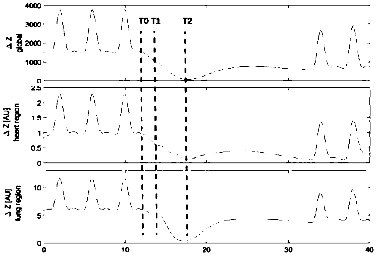
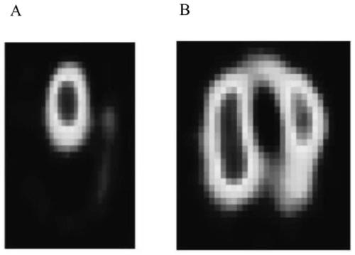
[0056] Example Construction and Joint Analysis of Lung Ventilation / Blood Flow Distribution Image

[0057] Measurement Cases and Data Acquisition

[0058] This study was approved by the institutional ethics committee. The inclusion criteria were patients with clinical diagnosis of respiratory failure, and established central venous catheter injection drug therapy. Exclusion criteria chest deformity or contraindication to electrical impedance monitoring (localized skin damage).

[0059] Patient information: 33 females, 50 males, average age 62 years old, most of them are patients with respiratory failure.

[0060] EIT raw data were obtained by PulmoVista 500 ( Lübeck, Germany), the EIT electrode belt containing 16 sensing electrodes was placed in the fourth to sixth intercostal spaces of the patient, and the reference electrode was placed in the abdomen. The current excitation mode is adjacent excitation mode, a single EIT image contains 32×32 pixels, and contains 20 frames...


PUM


Abstract

The invention discloses a bedside pulmonary ventilation-blood perfusion impedance tomography method based on saline radiography, an image monitoring device based on the method, an image monitoring system based on the method and a pulmonary embolism diagnosis system based on the method. By means of the method, the quality of blood perfusion imaging is improved, and dead space ventilation%, intrapulmonary shunt% and regional ventilation-blood match% are calculated according to blood perfusion imaging, so that the practicability is higher. Pulmonary embolism can be diagnosed by means of the method, the sensitivity is 90.9%, the specificity is 98.6%, and the method has very high clinical application value.

Description

technical field [0001] The invention belongs to the field of clinical medicine, and in particular relates to a bedside electrical impedance tomography method based on saline contrast-enhanced lung ventilation-blood perfusion. Background technique [0002] Electrical impedance tomography (EIT) is a non-invasive imaging technique used to study and monitor regional lung ventilation and perfusion (blood flow) in humans and animals. In contrast to conventional methods, EIT does not require the patient to breathe through a tube or sensor, does not apply ionizing X-rays, and can be used for extended periods of time, such as 24 hours or even longer. Therefore, EIT can be used continuously and thus is suitable for real-time and long-term monitoring of therapeutic effects. EIT was first used to monitor respiratory function in 1983 and remains the only bedside method that allows continuous, non-invasive detection of regional changes in lung volumes, blood flow, and cardiac activity. ...

Claims


Application Information

Patent Timeline
Patent Type & Authority Applications(China)
IPC IPC(8): A61B5/053A61B5/08A61B5/085A61B5/00
CPCA61B5/0536A61B5/0809A61B5/085A61B5/72A61B5/7282
Inventor 何怀武隆云招展奇池熠袁思依
Owner PEKING UNION MEDICAL COLLEGE HOSPITAL CHINESE ACAD OF MEDICAL SCI
Why Eureka
Try Eureka