Adjustable blood vessel clamping device for 3D printing animal experiments

An animal experiment, 3D printing technology, applied in the field of biomedical engineering, to improve the quality of surgery, speed up surgery, improve efficiency and success rate

Pending Publication Date: 2020-07-28
SHANGHAI TONGJI HOSPITAL
View PDF0 Cites 0 Cited by
  • Summary
  • Abstract
  • Description
  • Claims
  • Application Information

AI Technical Summary

Problems solved by technology

[0006] There is no relevant report about the adjustable blood vessel clamping device for 3D printing animal experiments of the present invention.

Method used


Image

  • Adjustable blood vessel clamping device for 3D printing animal experiments
  • Adjustable blood vessel clamping device for 3D printing animal experiments
  • Adjustable blood vessel clamping device for 3D printing animal experiments

Examples


Embodiment 1

[0059] Example 1 3D printing adjustable blood vessel clamping device for animal experiments

[0060] Please refer to attached Figure 1-6 .

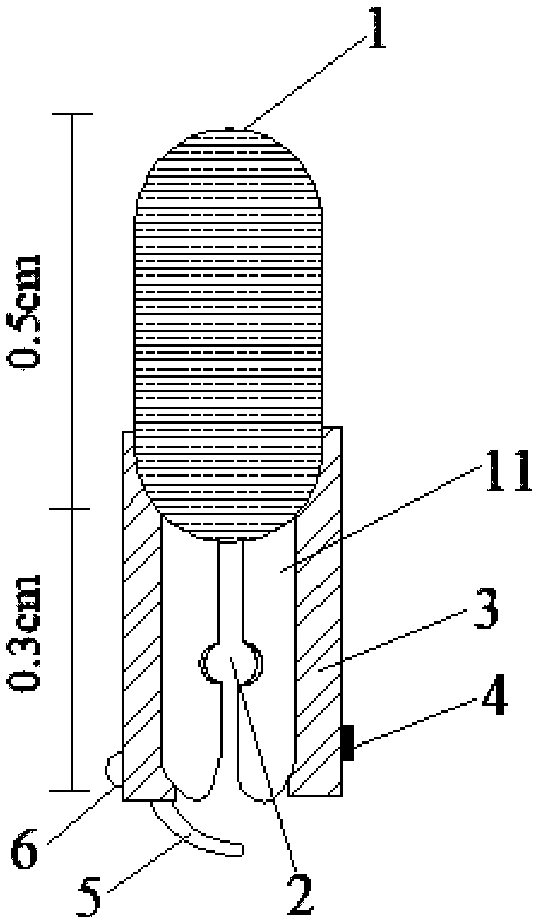
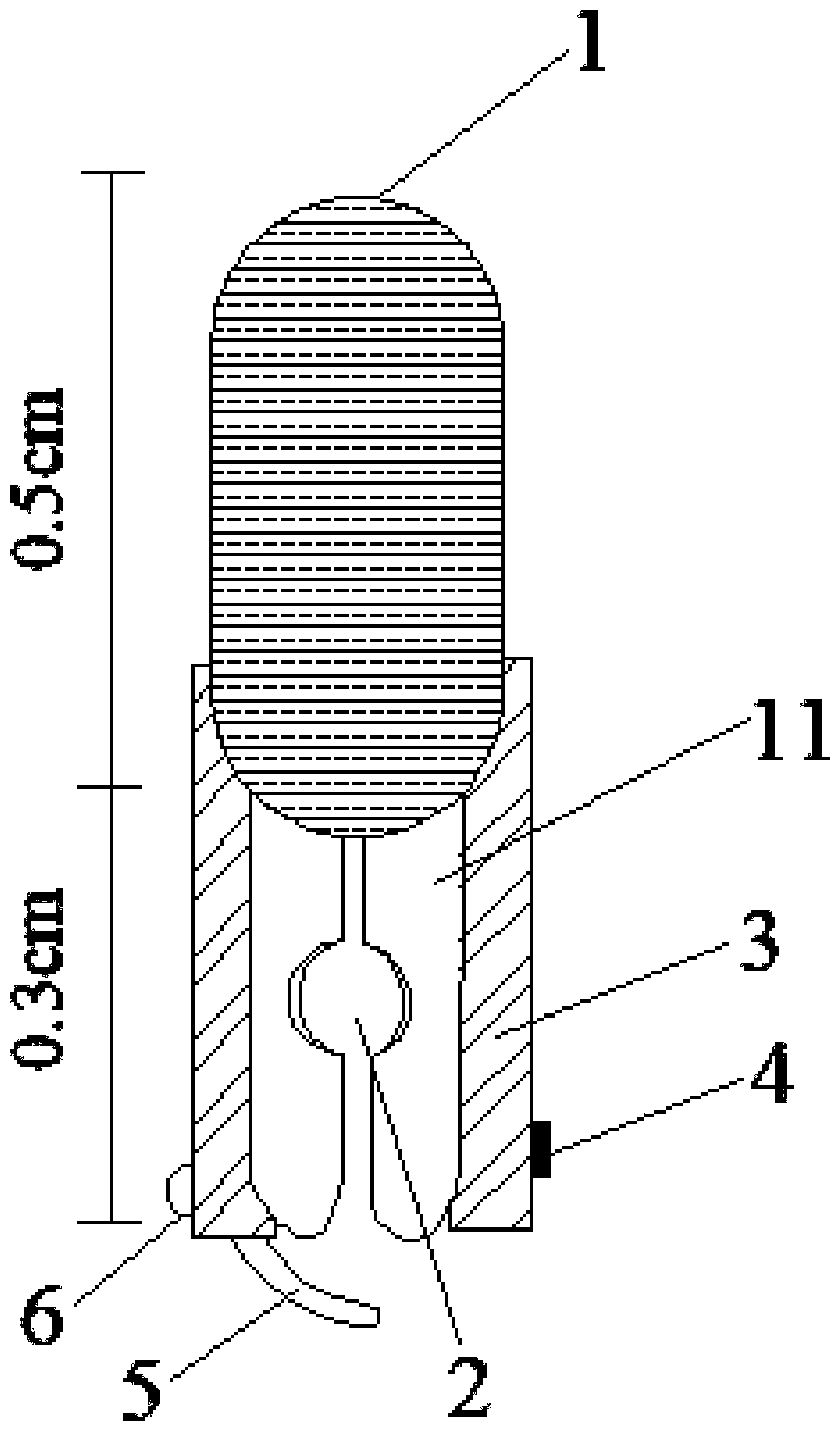
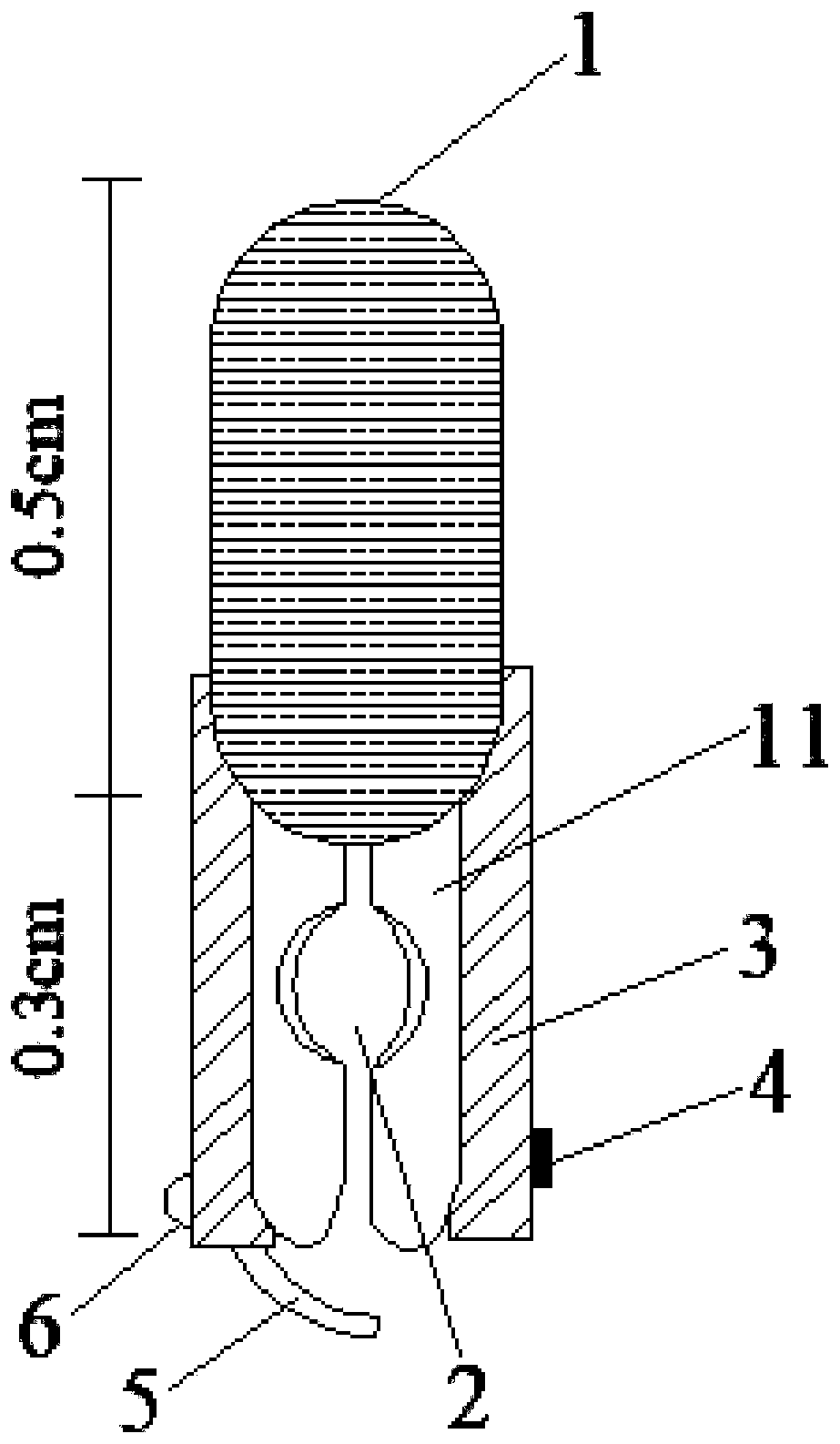
[0061] attached figure 1 It is a schematic diagram of the front structure of the small-diameter clamping hole of the present invention. attached figure 2 It is a schematic diagram of the front structure of the middle-diameter clamping hole of the present invention. attached image 3 It is a schematic diagram of the front structure of the large-aperture clamping hole of the present invention. attached Figure 4 It is a side structure schematic diagram of the present invention. attached Figure 5 It is a schematic diagram of the rubber anti-slip structure of the forearm of the vascular clamp. attached Image 6 It is a schematic diagram of the resin layer structure of the vascular clip.

[0062] An adjustable blood vessel clamping device for 3D printing animal experiments, including a blood vessel clamp body, a blocking structur...

Embodiment 2

[0073] Embodiment 2 Comparison of two different blood vessel clips

[0074] See Figure 7 , A. Current status of vascular clip: I. Physical map of vascular clip; II. Physical map of smooth vascular clip forearm; III. Schematic diagram of vascular clip, with a total length of about 1.6cm; IV. Schematic diagram of vascular clip forearm; B. 3D printing can regulate blood vessels Schematic diagram of the clamp, the length of the vascular clamp body is 0.5cm, and the length of the vascular clamp arm is 0.3cm. 1, 2, and 3 are schematic diagrams of vascular clips with mild, moderate, and severe stenosis.

Embodiment 3

[0075] Embodiment 3 Establishment of Chronic Cerebral Ischemia Animal Model

[0076] 1 Experimental method

[0077] 1) Pentobarbital sodium (4mg / kg) with a mass fraction of 2% anesthetizes the rat;

[0078] 2) Rats were fixed in the supine position, and after routine disinfection, a midline incision was made on the neck. like Figure 8 As shown in A, carefully separate the muscles, blood vessels and mucous membranes to expose the bilateral common carotid arteries without damaging the peripheral nerves;

[0079] 3) The right common carotid artery was ligated with 4-0 surgical thread, and the bilateral common carotid artery was ligated in the model group (carotid artery ligation);

[0080] 4) The vascular clip clamped the left common carotid artery, and the model group (vascular clamp) clamped the bilateral common carotid artery;

[0081] 5) Suture the skin layer by layer;

[0082] 6) The normal group underwent the same operation, but without ligation or clamping of blood v...


PUM


Abstract

The invention relates to an adjustable blood vessel clamping device for 3D printing animal experiments. The adjustable blood vessel clamping device comprises a body, stop blocks, a fixing structure, an anti-skid structure and a layered structure, wherein the blood vessel clamping body comprises a blood vessel clamping handle, a clamping hole and blood vessel clamping front arms; the fixing structure comprises a first protrusion, an elastic rope and a second protrusion; the anti-skid structure comprises anti-skid rubber; the layered structure comprises a first resin layer, a second resin layerand a third resin layer; the blood vessel clamping front arms are connected with the blood vessel clamping handle; the clamping hole is formed between the two blood vessel clamping front arms; the inner surfaces of the two blood vessel clamping front arms are connected with the anti-skid rubber in an adhesive mode; the outer surfaces of the two blood vessel clamping front arms are connected with the stop blocks; the elastic rope is located below the left stop block; the first protrusion is located on the side face of the right stop block; and the second protrusion is located on the side face of the left stop block. The reason of the high death rate of an animal model is considered, the chronic process of clinical cerebral ischemia from morning to evening and from light to heavy is simulated from the angle that clamping arms are provided with holes with different diameters, and the defect of the high death rate is overcome.

Description

technical field [0001] The invention relates to the technical field of biomedical engineering, in particular to an adjustable blood vessel clamping device for 3D printing animal experiments. Background technique [0002] 3D printing technology is to group or fuse materials layer by layer through a computer-aided printer system, and print out fine-structured, highly simulated models or workpieces layer by layer. technology etc. In recent years, 3D printing technology, as a cutting-edge technology, has been widely used in industry, construction, biology, aerospace engineering and other fields, and its important role in the medical field has also attracted much attention. Using 3D printing technology, the solid model of the fracture can be printed out, which can be used for steel plate pre-bending and guide plate production; implanting oral incomplete teeth and dermal defects; constructing 3D anatomical organs, simulating complex surgical procedures, etc. Compared with tradit...

Claims


Application Information

Patent Timeline
Patent Type & Authority Applications(China)
IPC IPC(8): A61B17/122B33Y50/00B33Y80/00B33Y40/20
CPCA61B17/122B33Y40/00B33Y50/00B33Y80/00
Inventor 王大鹏海舰
Owner SHANGHAI TONGJI HOSPITAL
Why Eureka
Try Eureka