Method and device for detecting standard section image in fetal ultrasound image

An ultrasound image and section image technology, which is applied in the directions of ultrasound/acoustic/infrasound image/data processing, image enhancement, image analysis, etc. Can not keep up with other problems, achieve real-time accurate diagnosis information, reduce detection pressure, and speed up detection

Pending Publication Date: 2020-07-28
HUNAN UNIV
View PDF18 Cites 8 Cited by
  • Summary
  • Abstract
  • Description
  • Claims
  • Application Information

AI Technical Summary

Problems solved by technology

[0003] At present, in the process of doctor detection, intelligent ultrasound equipment can provide auxiliary functions for screening standard sections and measuring parameters. Usually, the overall detection algorithm is placed on the CPU or GPU for coarse-grained parallelism. In clinical applications, the calculation speed of CPU or GPU It cannot keep up with the speed of ultrasound equipment to generate picture frames, and cannot realize frame-by-frame processing, resulting in the loss of some detection sections, making it difficult to provide real-time and accurate diagnostic information
Therefore, the existing technology has the problem of low detection speed and efficiency

Method used


Image

  • Method and device for detecting standard section image in fetal ultrasound image
  • Method and device for detecting standard section image in fetal ultrasound image
  • Method and device for detecting standard section image in fetal ultrasound image

Examples


Embodiment Construction

[0042] In order to make the purpose, technical solution and advantages of the present application clearer, the present application will be further described in detail below in conjunction with the accompanying drawings and embodiments. It should be understood that the specific embodiments described here are only used to explain the present application, and are not intended to limit the present application.

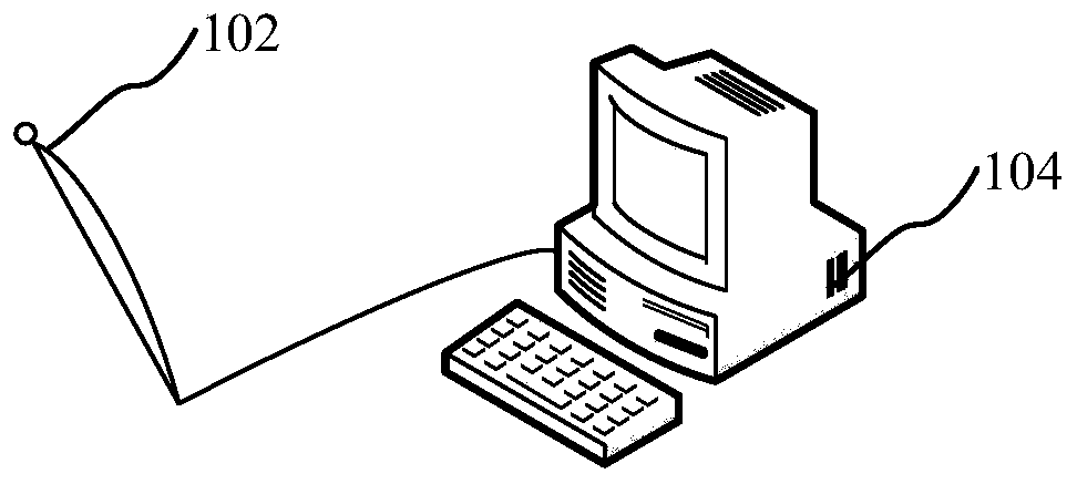
[0043] The detection method of the standard section image in the fetal ultrasound image provided by this application can be applied to such as figure 1The ultrasonic equipment shown includes an ultrasonic probe 102 and a processing terminal 104 connected to the ultrasonic probe 102. The processing terminal 104 has operating peripherals such as a display screen and a mouse and a keyboard, and also has multiple processors capable of parallel processing, The processor can be a CPU or a GPU. Wherein, the preprocessor of the processing terminal 104 acquires a preset number of ...


PUM


Abstract

The invention relates to a method and device for detecting a standard section image in a fetal ultrasound image, computer equipment and a storage medium. The method comprises the following steps: acquiring a continuous frame sequence of a preset number of fetal ultrasound image frames; sequentially carrying out preprocessing operation on the continuous frame sequence of the fetal ultrasound imageframes to obtain preprocessed image frames; sending the preprocessed image frame to a first processor for parallel processing, and inputting the preprocessed image frame into a trained segmentation model by the first processor to obtain a segmentation result of the preprocessed image frame; and sending the segmentation result to a second processor for parallel processing, and inputting the preprocessed image frame into the trained classification model by the second processor to obtain a final detection result of the fetal ultrasound image frame and display the final detection result. The method can improve the detection efficiency.

Description

technical field [0001] The present application relates to the technical field of prenatal ultrasound detection, in particular to a detection method, device, computer equipment and storage medium for standard section images in fetal ultrasound images. Background technique [0002] Ultrasound imaging has been widely used in pregnancy diagnosis due to its advantages of low consumption, real-time imaging and no radiation. In the process of using ultrasound imaging to diagnose the fetus, doctors need to obtain standard sections of various body parts of the fetus, such as ventricles, cerebral hemispheres, heart, etc., and then identify the standard sections, check the main anatomical structures through the standard sections, and then perform For further diagnosis and examination, analyze and diagnose according to the growth of the fetus and whether the anatomical structure of the fetus is abnormal. Therefore, the detection and identification of standard slices is very important f...

Claims


Application Information

Patent Timeline
Patent Type & Authority Applications(China)
IPC IPC(8): G06T7/00G06K9/00G06K9/34G16H50/20A61B8/08
CPCG06T7/0012G16H50/20A61B8/0866G06T2207/10132G06T2207/30044G06V20/46G06V10/267
Inventor 李肯立阳王东范欣欣朱宁波廖清李胜利
Owner HUNAN UNIV
Why Eureka
Try Eureka