
Automatic remote radiography surgical robot system

A surgical robot and robotic technology, applied in surgical robots, control/regulation systems, instruments, etc., can solve problems such as inability to effectively reduce radiation damage to doctors, surgical risks, and lack of information integration.

Active Publication Date: 2020-09-15
BEIJING TIANTAN HOSPITAL AFFILIATED TO CAPITAL MEDICAL UNIV
View PDF8 Cites 9 Cited by
  • Summary
  • Abstract
  • Description
  • Claims
  • Application Information

AI Technical Summary

Problems solved by technology

Due to the complexity of radiography surgery, doctors often need to be exposed to the X-ray environment for a long time. Even if they are equipped with lead-containing protective clothing, they cannot effectively reduce the radiation damage to doctors.
Although the existing technology has proposed some interventional surgery robot equipment for this situation, it is still impossible to form an unattended operating room.
In addition, the existing robot equipment for interventional surgery lacks the integration of relevant information such as patient video images, wound images, and patient sign monitoring during the operation.
It is often necessary to set up multiple display screens to display the working status information of different components during the operation. The imaging equipment can only display the image of the contrast machine. Paying attention to the information on the display screen will inevitably affect the doctor's operation accuracy and cause surgical risks

Method used


Image

  • Automatic remote radiography surgical robot system
  • Automatic remote radiography surgical robot system
  • Automatic remote radiography surgical robot system

Examples


Embodiment Construction

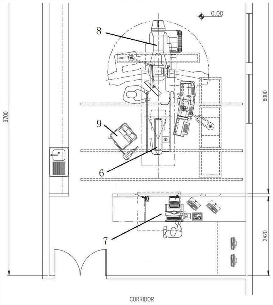
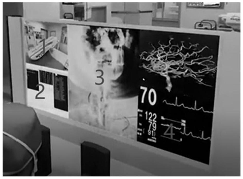
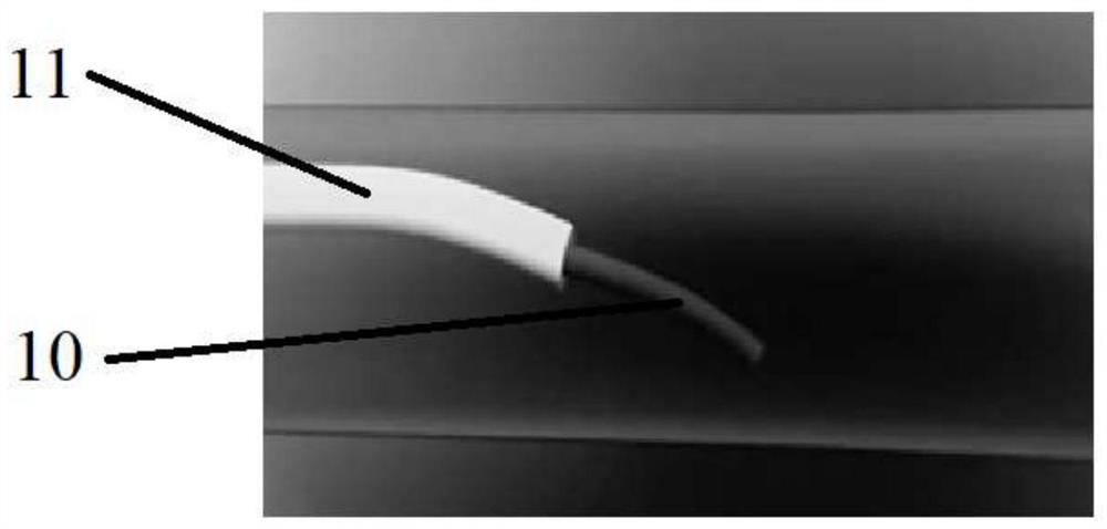
[0051] In order to explain the present invention better, so that understand, below in conjunction with appendix Figure 1-3 , the present invention is described in detail through specific embodiments.

[0052] Such as Figure 1-2 As shown, an automatic remote contrast surgery robot system proposed by the embodiment of the present invention includes: a robot 6, which is used for remote delivery of a guide wire catheter; a doctor console 7, which receives doctor instructions to Control the robot; DSA contrast machine 8, the DSA contrast machine generates contrast image information by emitting rays; high-pressure injector 9, the high-pressure injector is used to inject contrast medium; vital sign monitoring equipment, the vital sign monitoring The equipment is used to monitor the vital sign information 4 of the human body during the operation; the injection detector is set corresponding to the high-pressure injector to detect the injection parameters of the high-pressure injecto...


PUM


Abstract

The invention provides an automatic telegraphy surgical robot system. The system comprises a robot, a doctor console, a DSA radiography machine, a high-pressure injector, vital sign monitoring equipment, a injection detector, a parameter monitor and an information processing device; the robot is used for achieving remote delivery of a guidewire catheter; the doctor console is used for controllingthe robot by receiving instruction of a doctor; the DSA radiography machine is used for generating radiography image information by emitting rays; the high-pressure injector is used for injecting a contrast agent; the vital sign monitoring equipment is used for monitoring vital sign information of a human body in an operation process; the injection detector is used for detecting injection parameters of the high-pressure injector; the parameter monitor is arranged corresponding to the robot so as to monitor working state parameters of the robot; and the information processing device is used forprocessing the radiography image information, the human body vital sign information, the injection parameters and the working state parameters to generate first display data, and the first display data is displayed on a doctor console in a partitioned mode. The automatic remote radiography operation robot system can achieve an unattended operation room, and the harm of radiation to doctors in theradiography operation is reduced to the minimum.

Description

technical field [0001] The invention relates to the technical field of radiography surgery, in particular to an automatic remote radiography surgery robot system. Background technique [0002] Contrast surgery is a surgery that uses the imaging effect of contrast media to display visceral lesions. During the operation, doctors need to complete the operation with the help of X-ray-based digital silhouette angiography (DSA) guidance. Due to the complexity of radiography surgery, doctors often need to be exposed to the X-ray environment for a long time. Even if they are equipped with lead-containing protective clothing, they cannot effectively reduce the radiation damage to doctors. Although the prior art has proposed some interventional surgery robotic devices for this situation, it is still impossible to form an unattended operating room. In addition, the existing robot equipment for interventional surgery lacks the integration of relevant information such as patient video ...

Claims


Application Information

Patent Timeline
Patent Type & Authority Applications(China)
IPC IPC(8): A61B34/35A61M5/00A61M5/145A61M5/168A61B6/00A61B5/0205G16H40/67
CPCA61B5/02A61B5/02055A61B5/08A61B6/481A61M5/007A61M5/145A61M5/1684A61B34/35A61B2034/301G16H40/67
Inventor 李佑祥江裕华
Owner BEIJING TIANTAN HOSPITAL AFFILIATED TO CAPITAL MEDICAL UNIV
Learn More