Photoelectric chemical biosensor as well as preparation method and application of photoelectric chemical biosensor
A biosensor, photoelectrochemical technology, applied in the field of sensors, can solve the problems of limited photoelectric signal and sensitivity of photoelectric sensors, and achieve the effect of improving detection sensitivity
- Summary
- Abstract
- Description
- Claims
- Application Information
AI Technical Summary
Problems solved by technology
Method used
Image

Examples
Embodiment 1
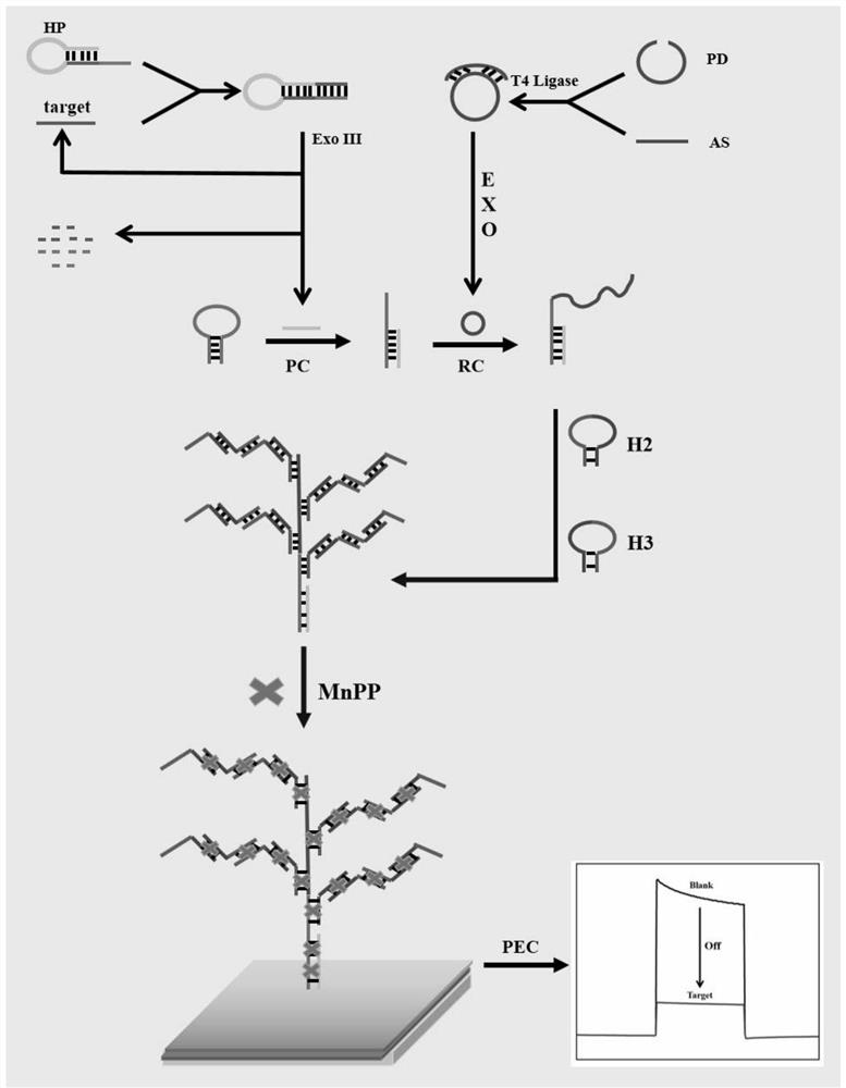
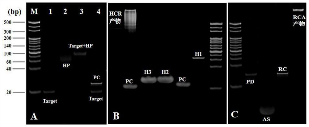
[0055] Mix 1 μL of padlock DNA with a concentration of 100 μM, 3 μL of assistantDNA with a concentration of 100 μM, 4 μL of secondary water and 3 μL of a buffer solution of 10×T4 DNA ligase, and anneal at 95°C for 5 min, then naturally cool to room temperature, and then add 120 U of T4DNA ligase was added to the mixed solution and connected at 37°C for 1h, then the mixed solution was reacted at 65°C for 10min to inactivate T4DNA ligase, after cooling to room temperature, add 4μL exonuclease buffer solution, 20U exonuclease I and 100U exonuclease III, cutting at 37°C for 2h, then reacting the mixed solution at 80°C for 15min to inactivate exonuclease I and exonuclease III, after cooling to room temperature, the DNA rolling circle template can be obtained , and store the obtained DNA rolling circle template at 4°C for future use.
[0056] Anneal 18 μL of HP with a concentration of 1 μM at 95°C for 5 minutes, then cool naturally to room temperature to obtain the hairpin structure...
Embodiment 2
[0059] Mix 1 μL of padlock DNA with a concentration of 100 μM, 3 μL of assistantDNA with a concentration of 100 μM, 4 μL of secondary water and 3 μL of a buffer solution of 10×T4 DNA ligase, and anneal at 97°C for 4 min, then naturally cool to room temperature, and then add 130 U of T4DNA ligase was added to the mixed solution and connected at 37°C for 2h, then the mixed solution was reacted at 68°C for 8min to inactivate T4DNA ligase, after cooling to room temperature, add 4μL exonuclease buffer solution, 30U exonuclease I and 110U exonuclease III, cutting at 37°C for 2h, then reacting the mixed solution at 83°C for 12min to inactivate exonuclease I and exonuclease III, after cooling to room temperature, the DNA rolling circle template can be obtained , and store the obtained DNA rolling circle template at 6°C for future use.
[0060] Anneal 18 μL of HP with a concentration of 1 μM at 97°C for 4 minutes, then cool naturally to room temperature to obtain the hairpin structure,...
Embodiment 3
[0063] Mix 1 μL of padlock DNA with a concentration of 100 μM, 3 μL of assistantDNA with a concentration of 100 μM, 4 μL of secondary water and 3 μL of a buffer solution of 10×T4 DNA ligase, and anneal at 93 °C for 6 min, then naturally cool to room temperature, and then add 1110 U of T4DNA ligase was added to the mixed solution and connected at 35°C for 1h, then the mixed solution was reacted at 62°C for 12min to inactivate T4DNA ligase, after cooling to room temperature, add 4μL exonuclease buffer solution, 10U exonuclease I and 90U exonuclease III, cutting at 35°C for 1h, then reacting the mixed solution at 77°C for 18min to inactivate exonuclease I and exonuclease III, after cooling to room temperature, the DNA rolling circle template can be obtained , and store the obtained DNA rolling circle template at 2°C for future use.
[0064] Anneal 18 μL of HP with a concentration of 1 μM at 93°C for 6 minutes, then cool naturally to room temperature to obtain the hairpin structur...
PUM
 Abstract
Description
Claims
Application Information
 - R&D
- Intellectual Property
- Life Sciences
- Materials
- Tech Scout
- Unparalleled Data Quality
- Higher Quality Content
- 60% Fewer Hallucinations
Browse by: Latest US Patents, China's latest patents, Technical Efficacy Thesaurus, Application Domain, Technology Topic, Popular Technical Reports.
© 2025 PatSnap. All rights reserved.Legal|Privacy policy|Modern Slavery Act Transparency Statement|Sitemap|About US| Contact US: help@patsnap.com