MRI hip joint inflammation segmentation and classification automatic quantitative grading sequential method

A quantitative grading, hip joint technology, applied in the field of image processing, can solve the problems of high consistency of voxel values ​​in the inflammatory area, affecting quantitative accuracy, poor quantitative results, etc., to improve the classification effect and classification effect, and improve the classification accuracy. , the effect of improving quantitative accuracy

Pending Publication Date: 2020-10-16
FOURTH MILITARY MEDICAL UNIVERSITY +1
View PDF0 Cites 6 Cited by
  • Summary
  • Abstract
  • Description
  • Claims
  • Application Information

AI Technical Summary

Problems solved by technology

In "Semi-automatic segmentation of JIA-induced inflammation in MRI images of ankle joints" published in "Progress in Biomedical Optics and Imaging" in 2019, a threshold-based inflammation segmentation method was proposed, using the prior information of MRI inflammation to set Thresholds are used to segment inflammation. This method has high requirements for the consistency of voxel values ​​in the inflammatory area. Due to the uneven distribution of voxels in the inflammatory area in MRI data, using this method will result in poor segmentation of the inflammatory area and affect the quantitative accuracy.
The method of lesion analysis based on the deep learning model UNet has been widely used in recent years. However, due t

Method used


Image

  • MRI hip joint inflammation segmentation and classification automatic quantitative grading sequential method
  • MRI hip joint inflammation segmentation and classification automatic quantitative grading sequential method
  • MRI hip joint inflammation segmentation and classification automatic quantitative grading sequential method

Examples


Embodiment 1

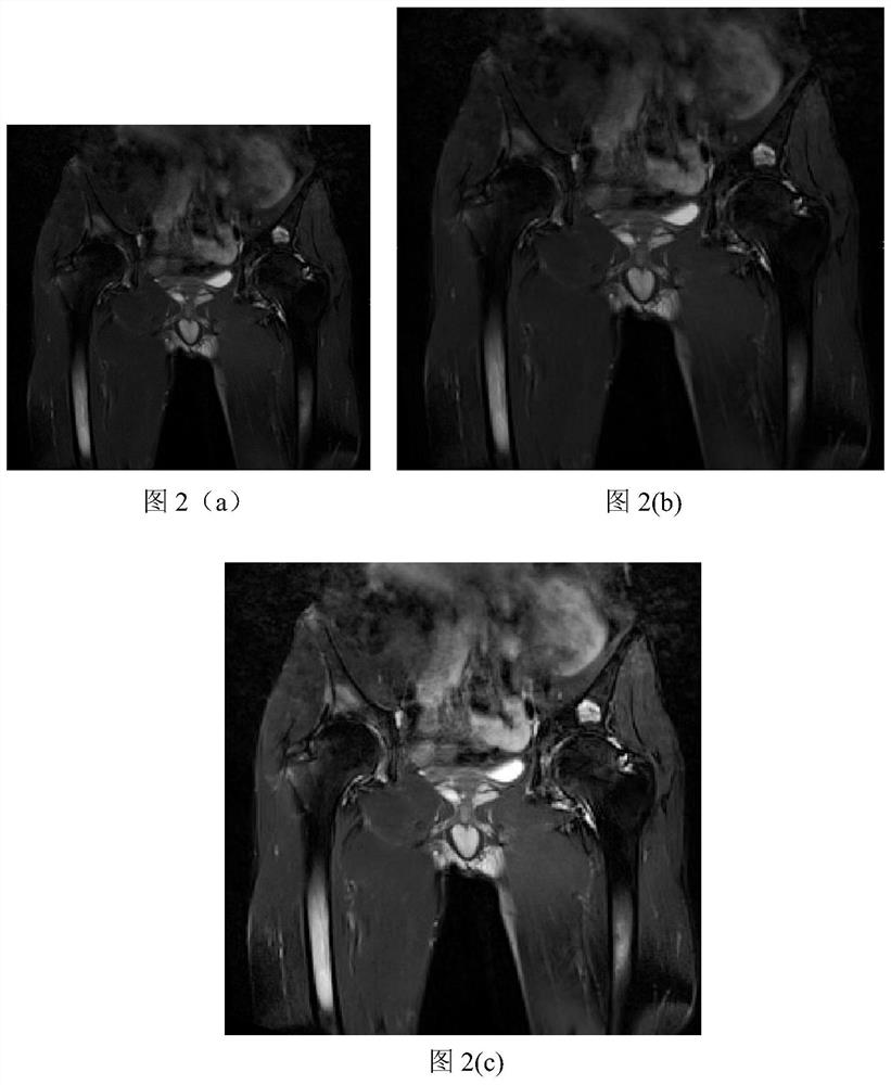
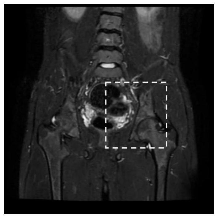
[0024] With the development of imaging technology, it provides a new way for doctors to understand the patient's illness. Doctors can combine imaging data to have a better understanding of spinal arthritis. Using MRI data, doctors can understand the disease of patients with spinal arthritis. However, when image processing algorithms are used to segment and classify MRI hip joint inflammation areas on MRI images, the shape and size of the MRI hip joint inflammation areas vary greatly, resulting in poor segmentation results. The MRI hip joint inflammation area as a whole The small proportion leads to poor classification of MRI hip inflammation. Aiming at the current situation, the present invention proposes an integrated automatic quantitative grading method combining segmentation and classification through thinking, exploration and experimentation, that is, MRI hip joint inflammation segmentation and classification automatic quantitative grading sequential method, which is used ...

Embodiment 2

[0039] The MRI hip joint inflammation segmentation and classification automatic quantitative grading sequential method is the same as that in Example 1, and the construction in step (3) of the present invention can automatically quantify the MRI hip joint inflammation area quantitative grading sequence based on segmentation and classification. The model is used for the automatic quantitative grading of multi-scale MRI hip joint inflammation areas, including the following steps:

[0040] (3.1) Constructing the segmentation classification model and multi-scale convolution module based on deep learning: firstly, using the mainstream segmentation network 3DResUNet of deep learning, classification network ResNet-50, and support vector machine, the segmentation classification model of MRI hip joint inflammation area was constructed. Aiming at the problem of large differences in the shape and size of inflammatory areas, the multi-scale convolution model GMS is composed of different vo...

Embodiment 3

[0048] MRI hip joint inflammation segmentation and classification automatic quantitative grading sequential method is the same as embodiment 1-2, the loss function of the two models described in the step (3.3) of the present invention, for the MRI hip joint inflammation area based on segmentation and classification Quantitative hierarchical sequential model training, which is expressed as follows:

[0049] Among them, the loss function L of the 3DResUNet part seg Expressed as follows:

[0050] L seg =L dice +λ*L wce

[0051]

[0052]

[0053] The loss function L of the segmentation model seg It is composed of two kinds of loss functions, which are the dice loss function L of the medical data segmentation network dice and weighted cross-entropy loss function L wce . Among them, C represents the maximum number of label categories, N represents the total number of pixels, c represents the category number, n represents the pixel number, p cn Indicates the probabilit...


PUM


Abstract

The invention discloses an MRI hip joint inflammation segmentation and classification automatic quantitative grading sequential method, solves the problem that hip joint inflammation areas are difficult to quantify and grade due to uneven distribution of shapes, sizes and voxel values of hip joints, and realizes automatic accurate quantification and grading evaluation of the hip joint inflammationareas. According to the method, the hip joint inflammation area label value is modified in an inverse exponential proportion according to the voxel value, so that the problem of poor segmentation result caused by large voxel value difference of the hip joint inflammation area is solved; according to the method, a multi-scale convolution module GMS is designed to improve the segmentation effect ofhip joint inflammation regions with large shape and size differences; aiming at the problem that hip joint inflammation regions with small proportions are difficult to classify, a segmentation modeland a classification model are fused to improve the classification precision. According to the method, integrated automatic quantitative grading of hip joint inflammatory regions with multiple scalesand large voxel value differences is realized, and the quantitative grading efficiency is improved. The method can be used for quantitative and graded automatic treatment of the MRI hip joint inflammation area.

Description

technical field [0001] The invention belongs to the technical field of image processing, and mainly relates to the segmentation and classification of nuclear magnetic resonance MRI hip joint inflammation lesions, specifically an MRI hip joint inflammation segmentation and classification automatic quantitative grading sequential method, which can be used in MRI images of hip joints in spinal arthritis Automated quantification and grading of inflammation. Background technique [0002] Spondyloarthritis is a chronic, inflammatory and disabling disease. Hip joint damage is the main cause of spondyloarthritis disability. The total annual incidence of total hip replacement is 0.17% to 5%. Therefore, accurate Early detection of hip joint damage in spondyloarthritis helps to assess the overall condition and formulate a diagnosis and treatment plan, thereby reducing the disability rate. Imaging methods have become the main method for early diagnosis and evaluation of spondyloarthrit...

Claims


Application Information

Patent Timeline
IPC IPC(8): G06T7/11G06K9/62G06N3/04G06N3/08
CPCG06T7/11G06N3/08G06T2207/10088G06T2207/30008G06N3/045G06F18/241
Inventor 韩青朱平黄陆光郑朝晖张葵丁进韩洁曹思颖缑水平
Owner FOURTH MILITARY MEDICAL UNIVERSITY
Why Eureka
Try Eureka