Visual distinguishing method for single-cell DNA epigenetic modification space positioning and adjacent distribution

A technology of epigenetic modification and spatial positioning, which is applied in the field of visual distinction between the spatial positioning and adjacent distribution of single-cell DNA epigenetic modification, and can solve the problems of identification interference and inability to obtain information on the spatial adjacent distribution of DNA epigenetic marks. , to achieve the effect of avoiding interference and high specificity

Active Publication Date: 2020-10-30
XI AN JIAOTONG UNIV
View PDF8 Cites 5 Cited by
  • Summary
  • Abstract
  • Description
  • Claims
  • Application Information

AI Technical Summary

Problems solved by technology

However, this method has not been applied in a real intracellular environment because the intracellular environment contains a large number of analogues of modified bases that interfere with recognition.
Therefore, current research methods cannot obtain information on the spatial proximity distribution of DNA epigenetic marks.

Method used


Image

  • Visual distinguishing method for single-cell DNA epigenetic modification space positioning and adjacent distribution
  • Visual distinguishing method for single-cell DNA epigenetic modification space positioning and adjacent distribution
  • Visual distinguishing method for single-cell DNA epigenetic modification space positioning and adjacent distribution

Examples


Embodiment 1

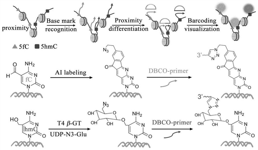
[0042] Example 1: Combination figure 1 , Visualization of the spatial location and neighboring distribution of DNA epigenetic modifications in MCF-10A cells. The cells were fixed on a cover glass with a micro reaction chamber using paraformaldehyde.

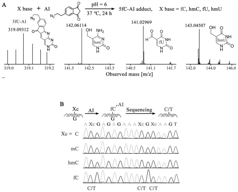
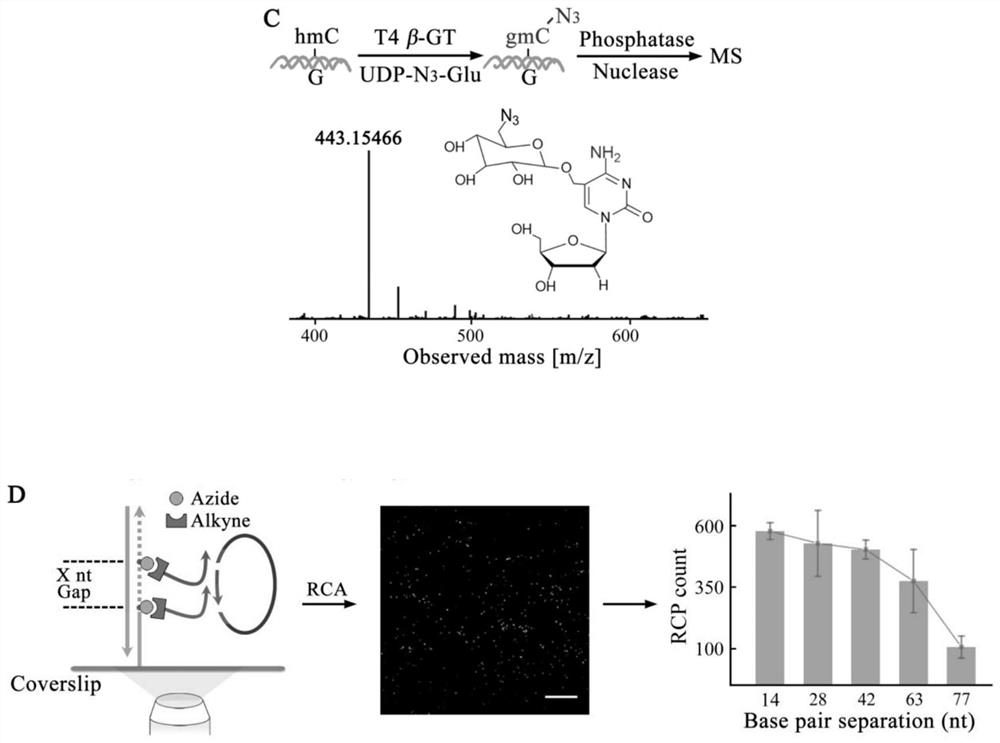
[0043] N of 5-fC by azide derivative probe AI 3 Chemical labeling of functional groups, the use of β-GT for 5-hmC N 3 Enzyme-mediated labeling of functional groups. 20 μL of 1×MES buffer containing 10 mM AI and 10% dimethyl sulfoxide was added to the reaction chamber and incubated at 37° C. for 24 h. Then, using click chemistry reaction, 1×PBS buffer containing 100 nM DBCO modified primer probe P1-fC was added to the reaction chamber, and the reaction was carried out at 37° C. for 1 h. Then add 20μL of UDP-N containing 1×NEBuffer 4, 50μM to the reaction chamber 3 -Glu and 5U T4β-GT culture solution, incubated at 37°C for 2h. Finally, 100nM P2-hmC probe was added to the reaction chamber and reacted at 37°C for 1h. Remove the react...


PUM


Abstract

The invention discloses a visual distinguishing method for single-cell DNA epigenetic modification space positioning and adjacent distribution, belongs to the field of analytical chemistry, and particularly relates to chemical labeling of an azide (N3) functional group on 5-formylcytosine (5-fC) through an azide derivative probe 1, 3-indadione (AI), and enzyme-mediated labeling of the azide (N3) functional group on 5-hydroxymethylcytosine (5-hmC) through T4 phage beta-glucosyltransferase (beta-GT). The paired primers close to the sites jointly achieve in-situ close connection on a ring template with a notch, the unpaired 5-fC and 5-hmC modification sites are left, the primers are released through strand displacement reaction, and then the corresponding specific ring template is hybridized;nucleic acid amplification and fluorescence probe hybridization are combined to realize single-cell DNA epigenetic modification space positioning and adjacently distributed single-molecule visual distinguishing.

Description

Technical field [0001] The invention belongs to the field of analytical chemistry and relates to a method for visually distinguishing the spatial positioning and neighboring distribution of single-cell DNA epigenetic modification. Background technique [0002] There are different DNA epigenetic marks in mammalian genomes. These epigenetic marks have a profound impact on gene expression and chromatin structure, and are related to the pathology of many diseases (such as cancer). Among the epigenetic markers of genes, 5-methylcytosine (5-mC) and its oxidized derivatives 5-hmC and 5-fC are mostly studied. After research, it is found that they are not only 5-mC demethylated It is an intermediate product of the basic pathway, and it also has good stability. Both of these DNA epigenetic markers are produced by the 10-11 translocation family of dioxygenases. [0003] The spatial location of epigenetic markers in a single cell is also the basis for understanding their functions. If diffe...

Claims


Application Information

Patent Timeline
Patent Type & Authority Applications(China)
IPC IPC(8): C12Q1/6853C12Q1/6841C12Q1/6806
CPCC12Q1/6853C12Q1/6841C12Q1/6806C12Q2531/125C12Q2563/107C12Q2565/601C12Q2523/109C12Q2531/119
Inventor 赵永席陈锋薛静张进
Owner XI AN JIAOTONG UNIV
Why Eureka
Try Eureka