Brain lesion area volume obtaining method and device based on deep learning, computer equipment and storage medium

A technology of deep learning and deep learning network, which is applied in computing, instruments for radiological diagnosis, medical science, etc. It can solve the problems of limited precision of prediction methods and poor extraction ability, and achieve the effect of improving calculation speed and accuracy

Pending Publication Date: 2021-03-02
HANGZHOU ARTERYFLOW TECH CO LTD
View PDF0 Cites 8 Cited by
  • Summary
  • Abstract
  • Description
  • Claims
  • Application Information

AI Technical Summary

Problems solved by technology

However, the non-threshold probability method has poor feature extraction abili

Method used


Image

  • Brain lesion area volume obtaining method and device based on deep learning, computer equipment and storage medium
  • Brain lesion area volume obtaining method and device based on deep learning, computer equipment and storage medium
  • Brain lesion area volume obtaining method and device based on deep learning, computer equipment and storage medium

Examples


Embodiment Construction

[0050] In order to make the purpose, technical solution and advantages of the present application clearer, the present application will be further described in detail below in conjunction with the accompanying drawings and embodiments. It should be understood that the specific embodiments described here are only used to explain the present application, and are not intended to limit the present application.

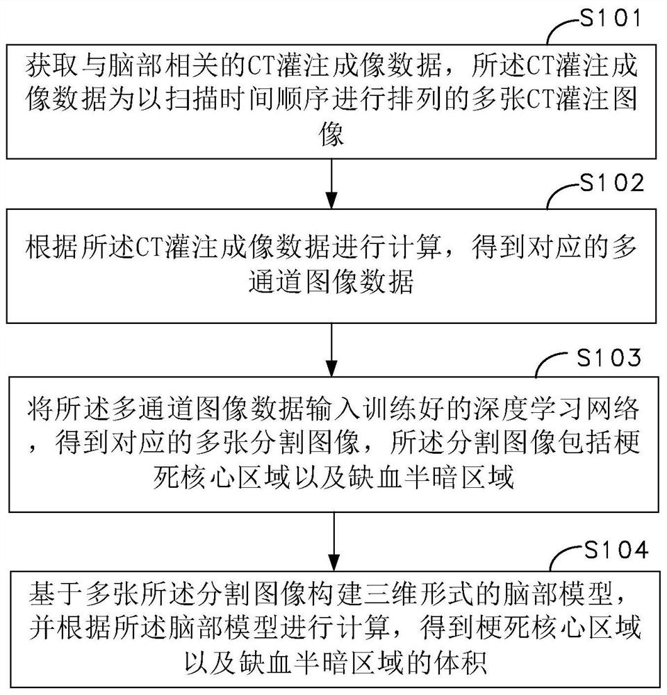
[0051] Such as figure 1 As shown, a method for obtaining the volume of brain lesion regions based on deep learning is provided, including the following steps:

[0052]Step S101, acquiring CT perfusion imaging data related to the brain, where the CT perfusion imaging data is a plurality of CT perfusion images arranged in the order of scanning time;

[0053] Step S102, performing calculations according to the CT perfusion imaging data to obtain corresponding multi-channel image data;

[0054] Step S103, input the multi-channel image data into the trained deep learning netw...


PUM


Abstract

The invention relates to a brain lesion area volume obtaining method and device based on deep learning, computer equipment and a storage medium. The method comprises the following steps: acquiring CTperfusion imaging data related to the brain, wherein the CT perfusion imaging data are a plurality of CT perfusion images arranged according to a scanning time sequence; calculating according to the CT perfusion imaging data to obtain corresponding multi-channel image data; inputting multi-channel image data into the trained deep learning network, and obtaining a plurality of corresponding segmented images, wherein each segmented image comprises an infarction core region and an ischemia semi-dark region; and constructing a brain model in a three-dimensional form based on the plurality of segmented images, and performing calculation according to the brain model to obtain volumes of the infarction core region and the ischemia semi-dark region. By adopting the method, the accuracy and the calculation speed can be improved.

Description

technical field [0001] This application relates to the interdisciplinary technical fields of image processing and medical engineering, and in particular to a method, device, computer equipment and storage medium for obtaining the volume of brain lesion regions based on deep learning. Background technique [0002] Stroke is also called stroke or cerebral infarction. Ischemic stroke is an acute cerebrovascular disease caused by insufficient blood supply to local brain tissue. Acute ischemic stroke is the most common type of stroke, accounting for about 60% of all strokes. ~80%, with a very high fatal risk. CTP (CT Perfusion) is a technical method of brain functional imaging. It reveals the physiological and pathological changes of brain diseases from the cellular level through the perfusion state of brain tissues and organs. It is of great value in judging the lesion area of ​​a patient and evaluating the infarct volume. [0003] After the onset of ischemic stroke, there will...

Claims


Application Information

Patent Timeline
IPC IPC(8): G06T7/00G06T7/62A61B6/00A61B6/03
CPCA61B6/032A61B6/481A61B6/501A61B6/5211A61B6/5217G06T7/0012G06T2207/10081G06T2207/30016G06T7/62
Inventor 鲁伟向建平
Owner HANGZHOU ARTERYFLOW TECH CO LTD
Why Eureka
Try Eureka