
Oval foramen unclosed detection method and system, terminal and storage medium

A detection method, foramen ovale technology, applied in image data processing, instruments, character and pattern recognition, etc., can solve the problems of not using artificial intelligence, high specificity, poor sensitivity, etc., to improve the detection rate and detection efficiency , simple operation and high accuracy

Active Publication Date: 2021-03-02
SHENZHEN INST OF ADVANCED TECH CHINESE ACAD OF SCI
View PDF8 Cites 6 Cited by
  • Summary
  • Abstract
  • Description
  • Claims
  • Application Information

AI Technical Summary

Problems solved by technology

[0007] 1. TEE is a semi-invasive detection method that cannot avoid risks. It is difficult to operate and requires high experience of clinicians. In China, this detection method is not popular, and only large hospitals usually carry out this detection; cTCD passed Cerebral blood circulation detection of contrast agent microbubbles from PFO can only infer the source of microbubbles indirectly, and cannot directly prove the existence of PFO; TTE is non-invasive and easy to operate, but its detection rate and sensitivity are low; cTTE as a A new type of detection method, which is non-invasive and can provide more detailed information on shunting from right to left in the heart, but is easily affected by noise, motion artifacts, and lung gas, with poor sensitivity and high specificity
[0008] 2. The existing clinical inquiry methods involve limited experimental groups and the conclusions are not universal, and their effects need to be further confirmed
[0009] 3. At present, there is no artificial intelligence method to effectively evaluate whether the foramen ovale is closed

Method used


Image

  • Oval foramen unclosed detection method and system, terminal and storage medium
  • Oval foramen unclosed detection method and system, terminal and storage medium
  • Oval foramen unclosed detection method and system, terminal and storage medium

Examples


Embodiment Construction

[0048] In order to make the purpose, technical solution and advantages of the present application clearer, the present application will be further described in detail below in conjunction with the accompanying drawings and embodiments. It should be understood that the specific embodiments described here are only used to explain the present application, not to limit the present application.

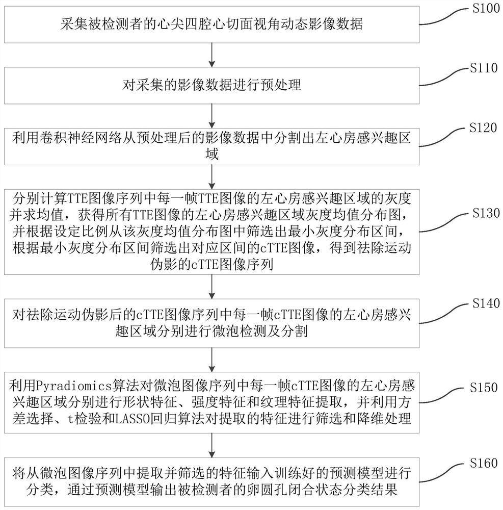
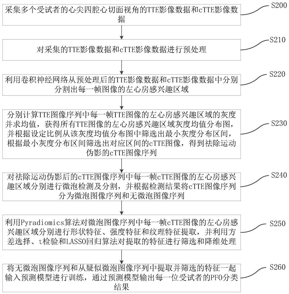
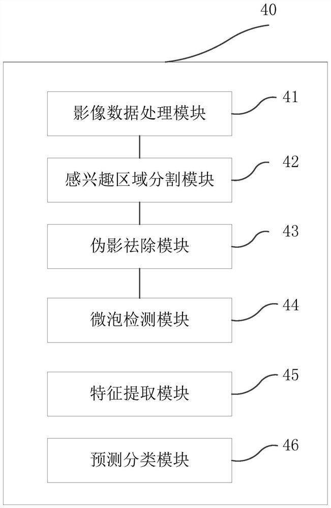
[0049] In view of the deficiencies in the prior art, the patent foramen ovale detection method of the embodiment of the present application collects the TTE image data and cTTE image data of the apical four-chamber section of the subject's heart respectively, and performs the TTE image data and the cTTE image data respectively. After the region of interest (ROI) of the left atrium was segmented, the cTTE image sequence was screened according to the minimum gray distribution interval of the ROI of the TTE image data to obtain the cTTE image sequence after removing motion artifacts. The scre...


PUM


Abstract

The invention relates to an oval foramen unclosed detection method and system, a terminal and a storage medium. The method comprises the following steps: respectively carrying out left atrium ROI segmentation on each frame of image in atrial apex four-cavity tangent plane view angle dynamic image data, wherein the cardiac apex four-cavity tangent plane view angle dynamic image data comprises TTE image data and cTTE image data in a set number of cardiac cycles; calculating the gray scale of the left atrium ROI of each frame of image in the TTE image data, screening out the minimum gray scale distribution interval of the TTE image data, and screening out the cTTE image of the corresponding interval in the cTTE image data according to the minimum gray scale distribution interval of the TTE image data; carrying out microbubble detection and feature extraction on the screened cTTE images, inputting the microbubble detection and feature extraction into a trained prediction model, and outputting an oval hole closing state detection result through the prediction model. Non-invasive PFO detection is realized, the operation is simple, the accuracy is high, and the detection rate and the detection efficiency are improved.

Description

technical field [0001] The application belongs to the technical field of medical ultrasound imaging, and in particular relates to a method, system, terminal and storage medium for detecting a patent foramen ovale. Background technique [0002] The foramen ovale is an embryonic defect (hole) in the atrial septum in the center of the heart. During fetal growth and development, the foramen ovale acts as an important life channel, which allows oxygenated blood to flow from the right atrium to the left atrium. In 75% of newborns, as the lungs develop and the resistance of the lungs decreases, there is a consequent increase in the pressure in the heart from the left to the right atrium, causing the opening to close automatically and the foramen ovale to functionally close . However, patent foramen ovale (PFO) still exists in 15%-35% of the adult population. According to statistics, PFO is the most common congenital heart abnormality among adults. The correct judgment of whethe...

Claims


Application Information

Patent Timeline
IPC IPC(8): G06T7/00G06T7/11G06T7/136G06K9/32
CPCG06T7/0012G06T7/11G06T7/136G06T2207/10132G06T2207/10016G06T2207/20081G06T2207/20084G06T2207/30048G06V10/25
Inventor 张世全肖杨杜丽娟马腾郑海荣
Owner SHENZHEN INST OF ADVANCED TECH CHINESE ACAD OF SCI
Learn More