Multi-modal image analysis method and system for cancer diagnosis

A cancer diagnosis, multi-modal technology, applied in image analysis, medical automation diagnosis, image enhancement and other directions, can solve the problems of poor clinical effect, low accuracy of breast cancer pathological classification evaluation, ignoring tumor heterogeneity, etc. Reduce missed diagnosis and misdiagnosis and improve screening efficiency

A cancer diagnosis, multi-modal technology, applied in image analysis, medical automation diagnosis, image enhancement and other directions, can solve the problems of poor clinical effect, low accuracy of breast cancer pathological classification evaluation, ignoring tumor heterogeneity, etc. Reduce missed diagnosis and misdiagnosis and improve screening efficiency

CN112489788APending Publication Date: 2021-03-12ZHONGNAN HOSPITAL OF WUHAN UNIV

Image

  • Multi-modal image analysis method and system for cancer diagnosis
  • Multi-modal image analysis method and system for cancer diagnosis
  • Multi-modal image analysis method and system for cancer diagnosis

Examples


Embodiment Construction

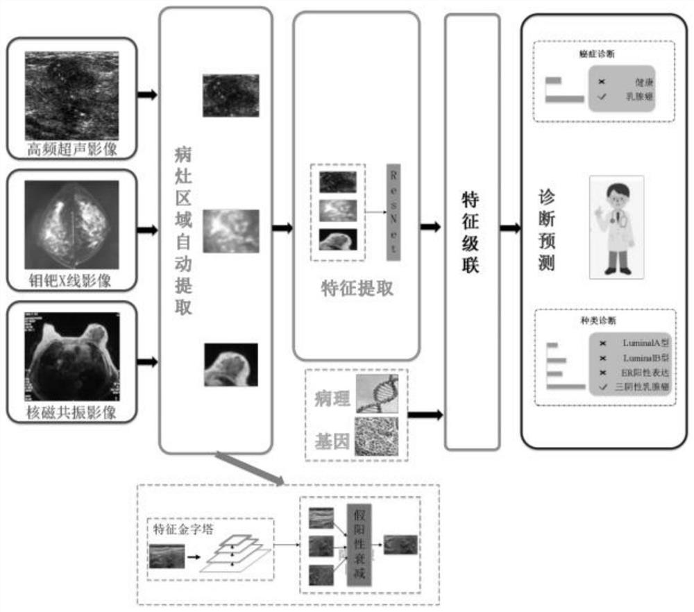
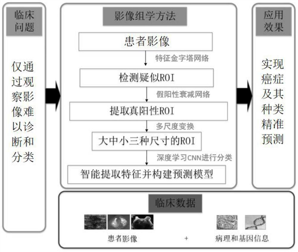
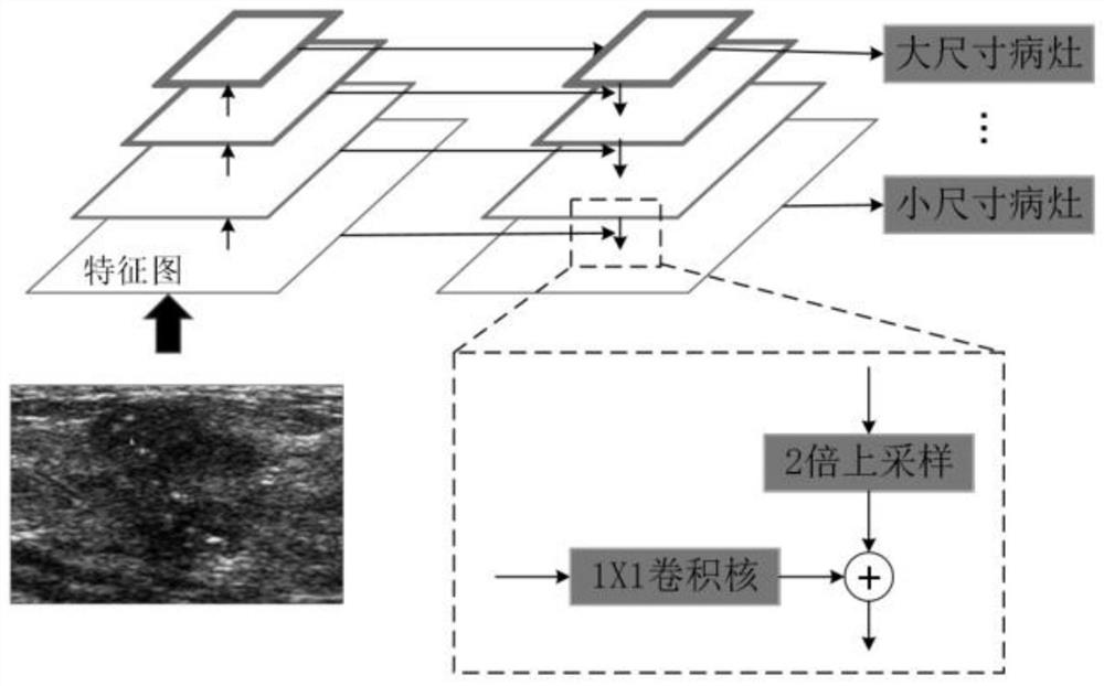
[0070] see figure 1 and figure 2 As shown, the embodiment of the present invention provides a multimodal image analysis method for cancer diagnosis, the method comprising the following steps:

[0071] Collect the image of the lesion area, and extract the suspected lesion area through the feature pyramid network;

[0072] The false positive attenuation network based on the context feature is used to classify the suspected lesion candidate area, and the true positive ROI is extracted;

[0073] The extracted true positive ROIs were multi-scale transformed and input into the integrated neural network, feature extraction and prediction model were constructed, combined with clinical and pathological information to predict the probability of pathological type, and output the weighted average of pathological type probability in the integrated neural network.

[0074] The method of the embodiment of the present invention can be applied to the diagnosis of breast cancer, thyroid canc...


PUM


Abstract

The invention provides a multi-modal image analysis method and system for cancer diagnosis, and relates to the technical field of image data analysis. The method comprises the steps: extracting a suspected lesion region through a feature pyramid network, carrying out the classification through a false positive attenuation network based on context features, and extracting a true positive ROI. According to the invention, the classification neural network based on the residual network is adopted, the pathological type is stably and accurately predicted, doctors are assisted in improving the screening efficiency, missed diagnosis and misdiagnosis are effectively reduced, and medical image-based cancer diagnosis and reasonable treatment of primary hospitals are facilitated.

Description

technical field [0001] The present invention relates to the technical field of image data analysis, in particular to a multimodal image analysis method and system for cancer diagnosis. Background technique [0002] Early cancer screening and treatment has become a major health issue worldwide, and imaging examinations are the main medical means for early detection and treatment of breast cancer, thyroid cancer and other cancers. Currently commonly used imaging techniques include mammography, ultrasound, MRI, etc., which can not only realize the location of lesions but also observe and evaluate the entire tumor tissue, such as location, number, size, shape, edge shape, density, internal echo, signal or degree of enhancement. Doctors use the observation of medical imaging pictures to determine the location and judgment of cancer foci and further evaluate the pathological types of tumors as much as possible. However, in the field of medical imaging, there is a large gap betwee...

Claims


Application Information

Patent Timeline
12 Mar 2021
Publication
CN112489788A
IPC
G16H50/20; G06T7/00; G06T7/11; G06K9/62; G06N3/04
CPC
G16H50/20; G06T7/0012; G06T7/11; G06T2207/10088; G06T2207/20084; G06T2207/30068; G06T2207/30096; G06N3/045
Inventors
吴猛