Method and device for fully convolutional single-stage breast image lesion detection based on multiple images

An image and breast technology, applied in the field of image processing, can solve the problems of low classification score, uneven shape distribution, long time, etc., to achieve the effect of improving the detection rate, improving sensitivity, and occupying less memory.

Active Publication Date: 2021-10-29
北京医准智能科技有限公司 +1
View PDF8 Cites 0 Cited by
  • Summary
  • Abstract
  • Description
  • Claims
  • Application Information

AI Technical Summary

Problems solved by technology

[0005] 1. The existing anchor-free detection methods generally only use the classification results located inside the object and the regression results of the rectangular frame of the object when identifying and regressing lesions, which will cause some low-quality prediction frames (and real The prediction frame with a large difference in the position of the rectangular frame) gets a higher classification score, while some high-quality prediction frames get a lower classification score, which further affects the subsequent non-maximum suppression process (NMS process, used to remove the non-maximum The result of the optimal prediction frame) makes the detection frame inaccurate
Moreover, the anchor-free method commonly used in the prior art is only aimed at mass lesions, and fails to cover all types and shapes of lesions, especially lesions with uneven shape distribution (such as calcification), thus resulting in missed detection
[0006] 2. The existing detection methods fail to make be...

Method used


Image

  • Method and device for fully convolutional single-stage breast image lesion detection based on multiple images
  • Method and device for fully convolutional single-stage breast image lesion detection based on multiple images
  • Method and device for fully convolutional single-stage breast image lesion detection based on multiple images

Examples


Embodiment 1

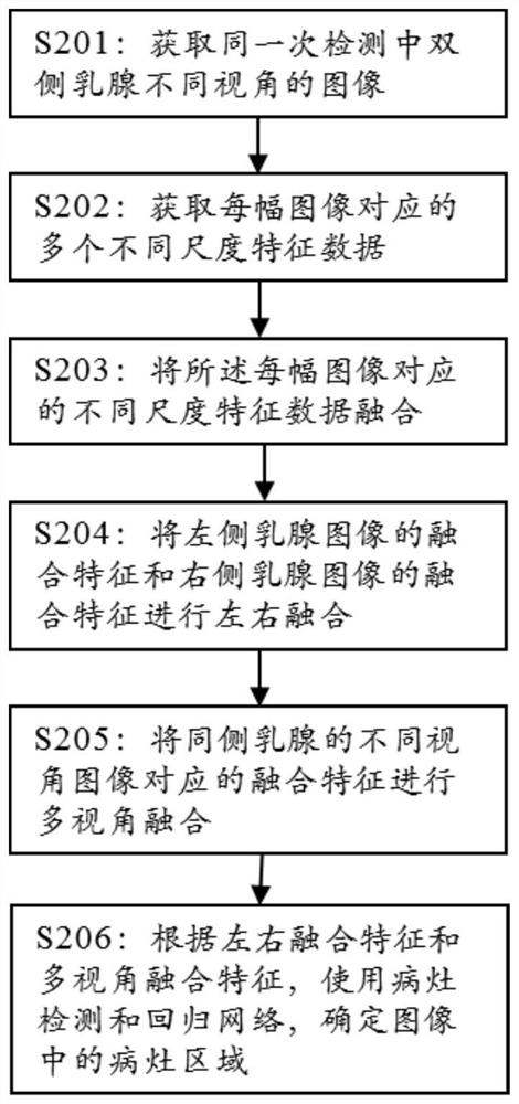
[0063] figure 2 A schematic block diagram of a breast image processing method according to an embodiment of the present invention is shown.

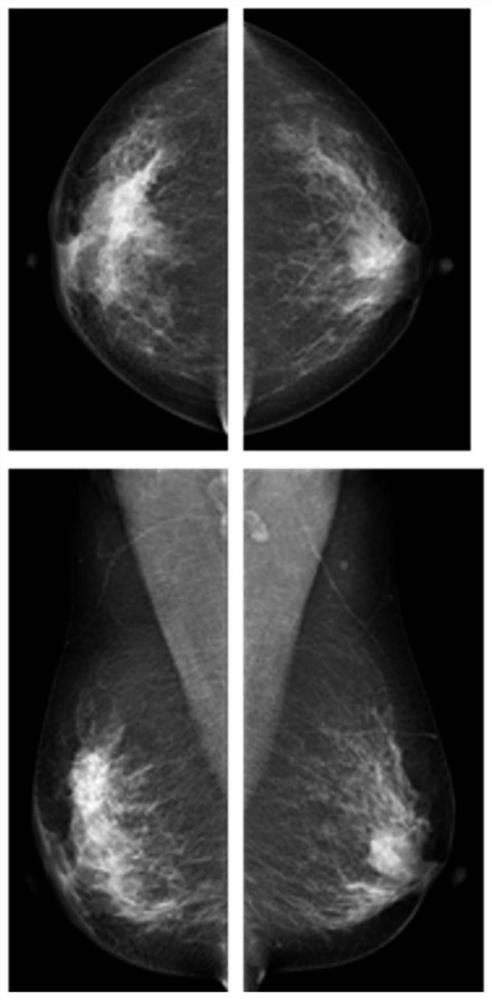
[0064] Such as figure 2 As shown, according to the processing method of mammary gland images according to an embodiment of the present invention, this embodiment takes mammograms as an example to illustrate the inventive solution, and the method of the present invention is not limited to mammograms (comprising ordinary X-ray images and It can also be used for images obtained by medical imaging methods such as color Doppler ultrasound, CT, and nuclear magnetic resonance used in breast examination.

[0065] A breast image processing method, comprising the following steps:

[0066] S201: Obtain images from different angles of view of the bilateral breasts in the same detection as images to be processed, wherein the images to be processed include: left craniocaudal (LCC) images, left mediolateral oblique (LMLO) images, right Lateral cra...

Embodiment 2

[0098] Such as Figure 9 As shown, Embodiment 2 of the present invention provides an image processing device, and the image processing device may be a computer program (including program code) running on a terminal. The image processing device can execute the breast image processing method in Embodiment 1, specifically including:

[0099] The image acquisition unit is used to acquire images of different angles of view of the bilateral mammary glands in the same detection as images to be processed, wherein the images to be processed include: left craniocaudal (LCC) images, left mediolateral oblique (LMLO) images ) image, right craniocaudal (RCC) image and right medial lateral oblique (RMLO) image;

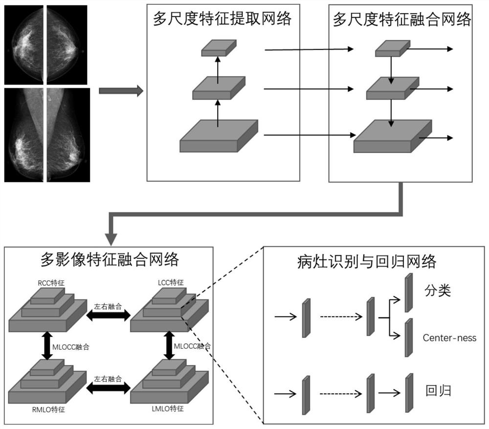
[0100] A multi-scale feature extraction unit is used to obtain a plurality of different scale feature data corresponding to each image to be processed;

[0101] A multi-scale feature fusion unit, configured to fuse a plurality of different-scale feature data corresponding to each ...

Embodiment 3

[0108] as attached Figure 10 The third embodiment of the present invention provides an electronic device, which is characterized in that it includes: a processor and a memory; the processor is connected to the memory, wherein the memory is used to store computer programs, and the processor is used to call The computer program is used to execute the breast image processing method described in Embodiment 1.

[0109] Electronic equipment in this embodiment may include but not limited to mobile phones, notebook computers, digital broadcast receivers, PDA (personal digital assistants), PAD (tablet computers), PMP (portable multimedia players), vehicle-mounted terminals (such as vehicle-mounted mobile terminals such as navigation terminals) and stationary terminals such as digital TVs, desktop computers, medical image acquisition devices, and the like. Figure 10 The illustrated terminal device is only an example, and should not limit the functions and application scope of the emb...


PUM


Abstract

Aiming at the fact that the existing breast lesion detection algorithm cannot well combine the information of bilateral breasts, cannot simultaneously meet the needs of identification and detection of multiple diseases including lumps and calcifications, and has a general effect on asymmetric dense glands, the present invention proposes A fully convolutional single-stage mammography lesion detection method with fusion of multi-image information. Use a non-anchor-based method for lesion detection, extract features of different scales from the original image, fuse the features of different scales, and fuse the information of different images, and finally directly predict whether a point on the feature map corresponds to a lesion and the specific location of the lesion.

Description

technical field [0001] The present invention relates to the field of image processing, in particular to a processing method for detecting lesion regions in mammary gland images. Background technique [0002] Breast cancer is the malignant tumor with the highest incidence rate in women. There are more than 270,000 new breast cancer cases in my country every year, and the incidence of breast cancer is increasing year by year, seriously threatening women's health. Early diagnosis of breast cancer is very important, early accurate diagnosis can increase the 5-year survival rate of breast cancer patients from 25% to 99%. Screening mammography is considered the test of choice for breast cancer screening. At present, mammography relies on subjective diagnosis, and the overall accuracy rate is not high enough and is limited by the level of evaluators. Compared with the personal experience judgment of a medical expert, the artificial intelligence recognition algorithm may identify ...

Claims


Application Information

Patent Timeline
IPC IPC(8): G06T7/00G06T7/73G06K9/46G06K9/62
CPCG06T7/0012G06T7/73G06T2207/10081G06T2207/20081G06T2207/20084G06T2207/30068G06T2207/30096G06V10/44G06V10/464G06F18/24
Inventor 王逸川赵子威王子腾王立威孙应实胡阳丁佳吕晨翀
Owner 北京医准智能科技有限公司
Why Eureka
Try Eureka