
Novel microglial cell activation method

A microglia cell and mouse technology, applied in animal cells, pharmaceutical formulations, medical preparations containing active ingredients, etc., can solve unseen problems, achieve simple operation, lower cost and risk, and broad application prospects Effect

Pending Publication Date: 2021-05-25
SHENZHEN INST OF ADVANCED TECH CHINESE ACAD OF SCI
View PDF0 Cites 0 Cited by
  • Summary
  • Abstract
  • Description
  • Claims
  • Application Information

AI Technical Summary

Problems solved by technology

But there is no similar report or product

Method used


Image

  • Novel microglial cell activation method
  • Novel microglial cell activation method
  • Novel microglial cell activation method

Examples


Embodiment 1

[0024] Example 1 Construction of Cx3cr1CreER-Ai27 genotype double-transferred mice

[0025] Cx3cr1CreER transgenic mice and Ai27 transgenic mice were both constructed from the Jackson Laboratory of the United States. The age of the mice is 2-3 months, the body weight is 20-30g, and there is no restriction on male or female. The experimental mice were provided with sufficient water and food, and were raised in an environment with 12 hours of darkness and 12 hours of light, and the room temperature was controlled between 20-25°C. Select Cx3cr1CreER mice with normal growth and Ai27 gene mice for hybridization. After hybridization, cut the tip of the mouse tail to about 4mm, use an electric iron to stop the bleeding of the mouse, put the mouse tail in a 1.5ml Eppendorf tube, add 500μL mouse Tail digestion buffer and 4 μL of proteinase K were digested in a water bath at 60°C for 12 hours. After the digested tissue was shaken, it was placed in a centrifuge at 10,000 rpm for 6 minu...

Embodiment 2

[0026] Example 2 Verification of Microglia Activation

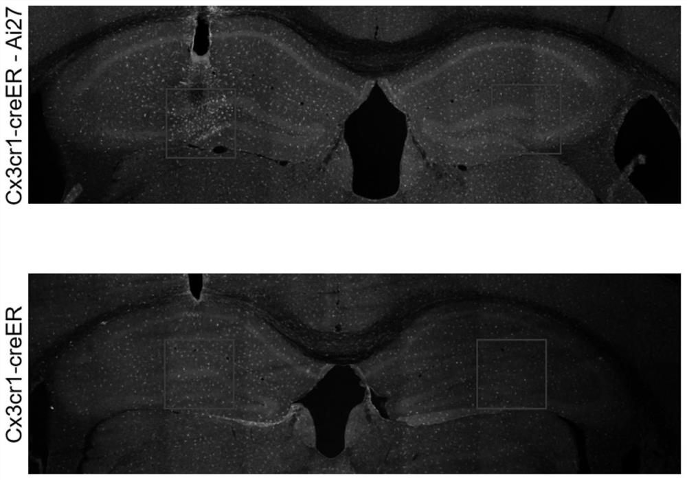
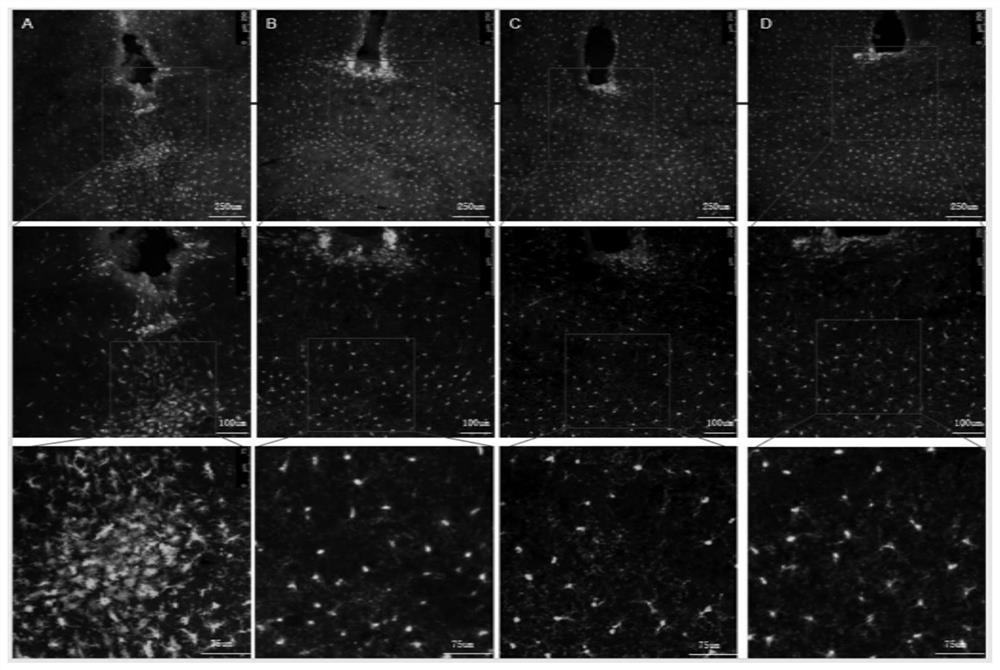
[0027]Referring to the construction method in Example 1, the Cx3cr1CreER-Ai27 genotype double transgenic mouse was obtained by crossing the Cx3cr1CreER transgenic mouse with the Ai27 transgenic mouse. In order to induce Cre recombinase, the Cx3cr1CreER-Ai27 genotype double transgenic mouse constructed in Example 1 The transgenic mice were stimulated with 4 mg Tamoxifen (purchased from Sigma) dissolved in 200 μl corn oil (Sigma), and Tamoxifen was injected subcutaneously or intraperitoneally at two time points at intervals of 48 hours. Cx3cr1CreER transgenic mice were used as the control group, and the same processing method.


PUM

PropertyMeasurementUnit
Wavelengthaaaaaaaaaa

Abstract

The invention provides a Cx3cr1CreER-Ai27 genotype double-transfected mouse and a novel microglial cell activation method. The Cx3cr1CreER-Ai27 genotype double-transfected mouse is obtained by hybridizing a Cx3cr1CreER-Ai27 genotype mouse and an Ai27 gene mouse, an optical fiber is implanted in a target brain region after Tamoxifen is injected into an abdominal cavity, and microglial cells in a region below the optical fiber can be specifically activated by using blue light (450-490nm) for stimulation. The microglial cell activation method can specifically activate the microglial cells and accurately distinguish the microglial cells from other cells, and has important significance in research on development, behaviors, functions and the like of the microglial cells, and the novel microglial cell activation method is simple and convenient to operate, reduces research cost and risks, and has wide application prospects.

Description

technical field [0001] The invention relates to the field of biology, in particular to a Cx3cr1CreER-Ai27 genotype double-transformed mouse, a new method for activating microglial cells and an application thereof. Background technique [0002] Microglia is the smallest glial cell in the central nervous system (CNS), distributed throughout the central nervous system, accounting for about 5%-10% of the total glial cells. As immune effector cells resident in the central nervous system, microglia belong to the mononuclear phagocyte family and are widely considered to be the main immune effectors of the central nervous system. The morphology of microglia is highly plastic, and its biological Functional status is closely related. In normal brain tissue, microglia are highly branched, with tertiary and quaternary branch structures, and the branches between cells rarely overlap. Branching microglia are often referred to as "resting microglia". Normally, highly branched resting-st...

Claims


Application Information

Patent Timeline
IPC IPC(8): A01K67/027
CPCA01K67/0275A01K67/0276A01K67/0278A01K2217/072A01K2217/075A01K2217/15A01K2227/105A01K2267/0312A01K2267/0318A01K67/027A61K35/30C12N5/06
Inventor 詹阳吕泽中
Owner SHENZHEN INST OF ADVANCED TECH CHINESE ACAD OF SCI
Learn More