Method and system for assisting in identifying submucosal blood vessels under endoscope

A blood vessel and mucous membrane technology, applied in the field of submucosal blood vessels under endoscopy, to ensure the safety of surgery and prolong the operation time

Active Publication Date: 2021-05-28
SHANDONG UNIV QILU HOSPITAL +1
View PDF7 Cites 2 Cited by
  • Summary
  • Abstract
  • Description
  • Claims
  • Application Information

AI Technical Summary

Problems solved by technology

There is currently no endoscopically assisted identification of submucosal vessels

Method used


Image

  • Method and system for assisting in identifying submucosal blood vessels under endoscope
  • Method and system for assisting in identifying submucosal blood vessels under endoscope
  • Method and system for assisting in identifying submucosal blood vessels under endoscope

Examples


Embodiment 1

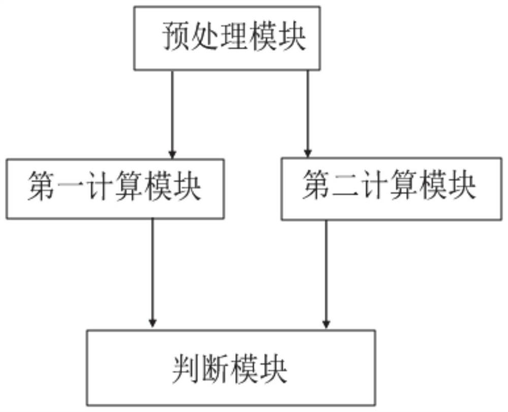
[0046]Such asfigure 1 As shown in the present invention, the present invention provides an inner mirror to assist in identifying a system of blood vessels under mucosa, the system comprising: a pretreatment module for pre-processing a timing image of the real-time acquired portion to be detected, timing image The pixel value is converted to zero value and unit variance;

[0047]The first computing module is used to extract blood volume waves from the pre-treated timing image based on the imaging photocatalytic capacitance, and determine the corresponding blood volume fluctuation frequency;

[0048]The second computing module is used to extract the pixel change value of each pixel point from the pre-treated timing image, and determine the pixel fluctuation frequency of the corresponding pixel points from the pre-treated timing image.

[0049]The determination module determines the actual vascular coverage area under the mucosa according to the blood volume wave, the blood volume wave frequenc...

Embodiment 2

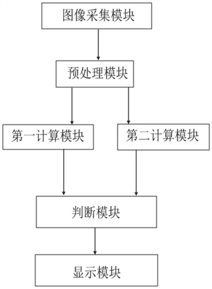
[0073]Such asfigure 2 As shown, the present invention provides an inner mirror to assist in identifying an intramidal blood vessel under mucosa, including:

[0074]Image acquisition module is used to obtain a timing image to be detected from the endoscopic check;

[0075]The pretreatment module is used to prepare the timing image of the real-time acquired site to be detected, and the pixel value of the timing image is converted to zero value and unit variance;

[0076]The first computing module is used to extract blood volume waves from the pre-treated timing image based on the imaging photocatalytic capacitance, and determine the corresponding blood volume fluctuation frequency;

[0077]The second computing module is used to extract the pixel change value of each pixel point from the pre-treated timing image, and determine the pixel fluctuation frequency of the corresponding pixel points from the pre-treated timing image.

[0078]The determination module is used to determine the actual vascular c...

Embodiment 3

[0105]Embodiment 3 of the present invention provides an inner mirror to assist in identifying a blood vessel under mucosa, including the following steps:

[0106]Step 1: Get the timing image of the detection site from the endoscopy

[0107]Keep the lens stable 2-3s using an endoscopic camera 2-3S to obtain an image of the N-frame RGB. During this process, the sensation of the perceptual hash algorithm is used to calculate the lens sway. When the two frame image hash value difference is greater than a certain threshold, the image information needs to be recording the image information.

[0108]Step 2: Preprocessing the collected image

[0109]The green component G image data is selected from the read RGB image data, as the original data of the extracted digestive blood flow information image. The reason is that the oxygen-containing hemoglobin has the greatest absorption rate of green light, and the experiment is compared to the maximum amount of pixel value caused by the blood of the G-channel ...


PUM


Abstract

The invention provides a method and system for assisting in recognizing submucosal blood vessels under an endoscope, and belongs to the technical field of blood vessel recognition. The method comprises the steps that a time sequence image, collected in real time, of a to-be-detected part is preprocessed, and the pixel value of the time sequence image is converted into a zero mean value and a unit variance; based on the imaging type photoplethysmography technology, blood volume waves are extracted from the preprocessed time sequence images, and the corresponding blood volume fluctuation frequency is determined; based on an imaging type photoplethysmography technology, a pixel change value of each pixel point is extracted from the preprocessed time sequence image, and a pixel fluctuation frequency of the corresponding pixel point is determined; and according to the blood volume wave, the blood volume fluctuation frequency, the pixel change value and the pixel fluctuation frequency, the submucosal blood vessel coverage area is determined. According to the method, in the operation process of the endoscope, on the premise that no extra equipment is used and the operation time is not prolonged, alimentary canal blood flow information can be accurately extracted in real time, submucosal blood vessels can be accurately recognized, and operation safety is guaranteed.

Description

Technical field[0001]The present invention relates to the field of vascular identification, and more particularly to an inner mirror auxiliary recognition method and system of blood vessels under mucosa.Background technique[0002]With the development of endoscopy, more and more diseases that need surgery, the disease can be selected, and the internal mirror micro-creation treatment can be selected. However, during the operation, it is inevitable that the problem of bleeding in the operation is inevitable due to various reasons such as mucosa. The lightweight can be treated inner mirror, and the weight is subject to hemostasis, and very few patients will even lead to death. There is currently no method for assisting the blood vessels under mucosa under endoscopy. Imaging Photoelectric Capacity Sacking Technology (IPPG), allows smartphones to achieve non-contact measurement of heart rate, breathing rate, autonomic excitability, blood pressure, breathing rate, autonomic neurons, blood p...

Claims


Application Information

Patent Timeline
Patent Type & Authority Applications(China)
IPC IPC(8): A61B5/02A61B5/0295
CPCA61B5/0084A61B5/02A61B5/0295A61B5/72
Inventor 左秀丽李延青马铭骏李真邵学军杨晓云赖永航冯健
Owner SHANDONG UNIV QILU HOSPITAL
Why Eureka
Try Eureka