Biological 3D printing method for large tissues

A 3D printing and tissue technology, applied in the field of 3D bioprinting of large tissue, can solve the problems of flow channel effectiveness, cumbersome operation, structural collapse, etc., achieve good biocompatibility, ensure survival rate, and solve the effect of collapse

Active Publication Date: 2021-06-08
苏州永沁泉智能设备有限公司
View PDF3 Cites 7 Cited by
  • Summary
  • Abstract
  • Description
  • Claims
  • Application Information

AI Technical Summary

Problems solved by technology

Compared with the traditional printing process, the multi-functional coaxial biological structure manufacturing process based on the gel tubular unit provides a better printing unit idea on the printing unit during the printing process, but because the structural strength of the hollow tube is often not enough Support the entire structure, causing the structure to collapse and the effectiveness of the runners to be affected
In addition, the original printing method requires secondary perfusion of endothelial and other vascularized functional cells for the reconstruction of vascularization inside the printed tissue, which is relatively cumbersome.

Method used


Image

  • Biological 3D printing method for large tissues
  • Biological 3D printing method for large tissues
  • Biological 3D printing method for large tissues

Examples


Embodiment 1

[0040] refer to figure 1 As shown, the present embodiment provides a bio-3D printing method of a large tissue, comprising the following steps:

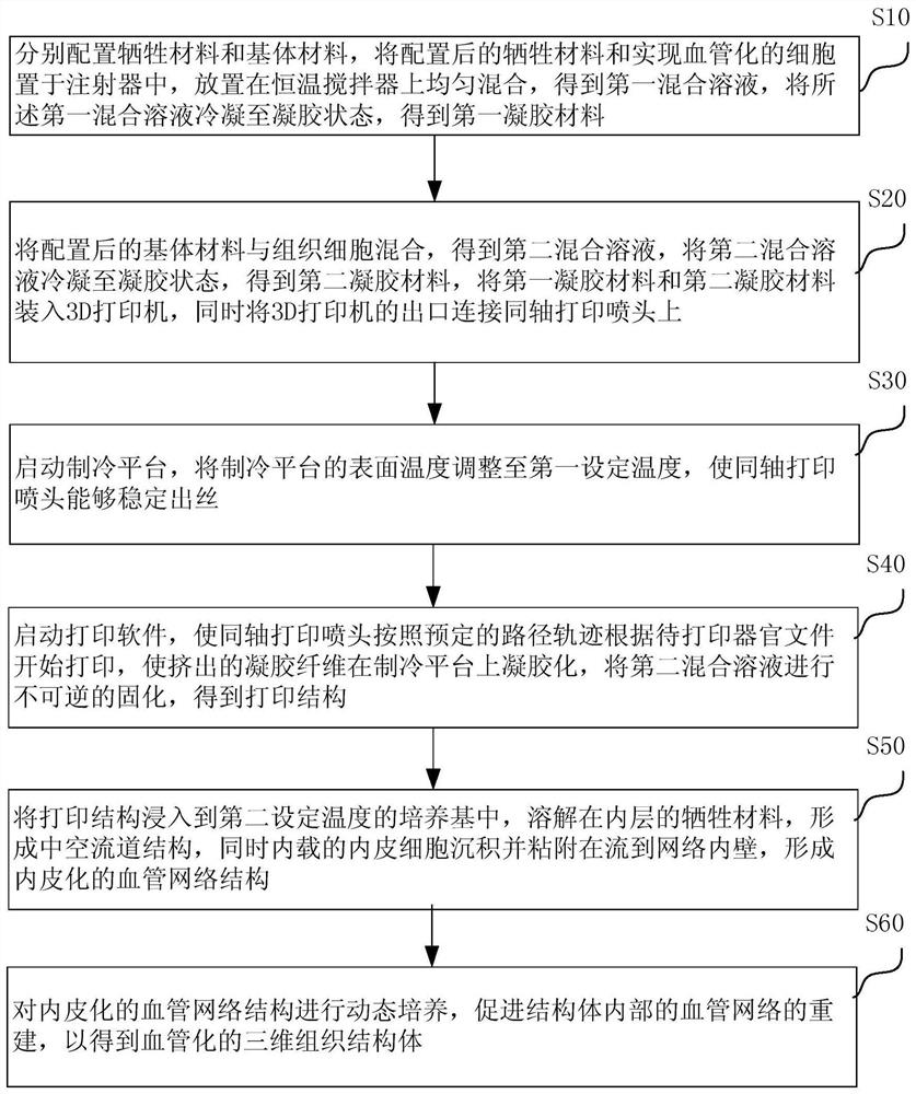
[0041] S10, prepare the sacrificial material and the matrix material respectively, put the prepared sacrificial material and cells that realize vascularization (such as endothelial cells, cells that can be induced to differentiate into endothelial tissue) into a syringe, and place them on a constant temperature stirrer for uniform mixing , to obtain a first mixed solution, and condense the first mixed solution to a gel state to obtain a first gel material.

[0042] The aforementioned sacrificial material may include gelatin and other materials with good biocompatibility, printability, and easy removal. The above-mentioned matrix material may include GelMA (methacrylic anhydride gelatin) and other materials with good biocompatibility, printability, stability and non-deformation characteristics.

[0043] Specifically, the sacrificial ...

Embodiment 2

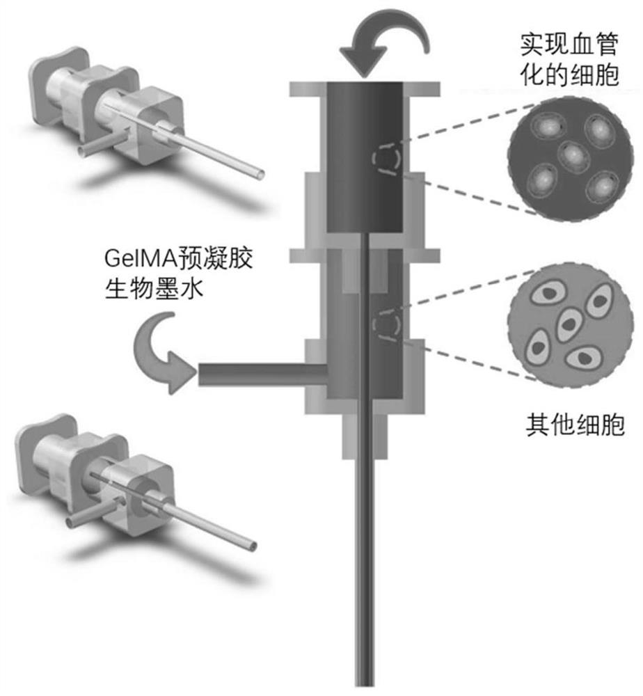
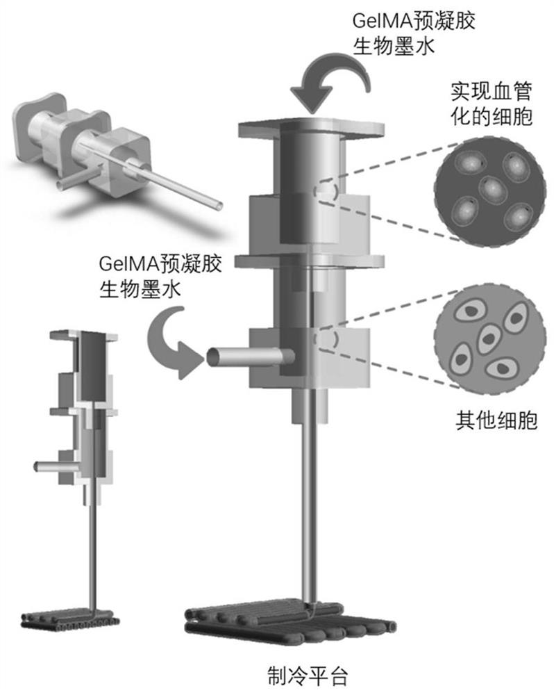
[0079] In this embodiment 2, the devices involved in the execution of the bio-3D printing method of the above-mentioned bulk tissue may include: a bio-3D printer with a motor module of the three coordinate axes of XYZ controlled; coaxial printing nozzles, external nozzles The inner diameter is up to 3mm, and the inner diameter of the inner nozzle is up to 0.5mm; a cooling platform with real-time temperature control; a syringe pump system with controllable flow rate.

[0080] Specifically, the device for realizing the bio-3D printing method of the above-mentioned bulk tissue may include: a printing system and a refrigeration system. The above printing system can refer to figure 2 As shown, the motor is an ordinary two-phase four-wire stepping motor, which passes through the motor base of the module coaxially. The two are fixedly connected by four screws, and the limit switch is fixed on the motor base of the module on the motor side by two screws. , used to realize positionin...


PUM


Abstract

The invention relates to a biological 3D printing method for large tissues. A prepared sacrificial material and vascularized cells are placed in a syringe to obtain a first mixed solution and a first gel material corresponding to the first mixed solution;a prepared matrix material is mixed with tissue cells to obtain a second mixed solution and a second gel material corresponding to the second mixed solution, the first gel material and the second gel material are loaded into a 3D printer, and an outlet of the 3D printer is connected to a coaxial printing spray head; the coaxial printing spray head starts printing according to a preset path track and according to a to-be-printed organ file, extruded gel fibers are gelatinized on a refrigeration platform, matrix material components in the gel fibers are irreversibly cured, and a printing structure is obtained; the printing structure is immersed in a culture medium at a second set temperature, a hollow flow channel structure is formed, and a network structure with vascular cells is obtained; and the vascular network structure is subjected to dynamic culture, reconstruction of a vascular network in the structure is promoted, and therefore a vascularized three-dimensional tissue structure capable of keeping effectiveness of a flow channel is obtained.

Description

technical field [0001] The invention relates to the technical field of biomanufacturing, in particular to a method for bio-3D printing of bulk tissues. Background technique [0002] In recent years, 3D printing technology has gradually been used in tissue engineering and regenerative medicine to solve the problem of lack of organ donors. The idea of ​​traditional tissue engineering technology is to plant the corresponding cells on the pre-printed biological scaffolds, and carry out in vitro simulation of in vivo culture or transplantation into the body. Compared with traditional tissue engineering, bio-3D printing technology mixes cells in bio-ink when printing tissues or organs. In this way, the cell density can be achieved, and the spatial distribution of multiple types of cells in different structural materials can be controlled. Therefore, bioprinting technology, also known as cell printing and organ printing, has gradually become a research hotspot in the manufacture o...

Claims


Application Information

Patent Timeline
Patent Type & Authority Applications(China)
IPC IPC(8): B29C64/106B29C64/379B29C64/40B33Y10/00B33Y40/00B33Y40/20B33Y70/10
CPCB29C64/106B29C64/379B29C64/40B33Y10/00B33Y40/00B33Y70/00
Inventor 贺永陈路路邵磊高庆
Owner 苏州永沁泉智能设备有限公司
Why Eureka
Try Eureka