
Preparation method of transmission electron microscope sample

A transmission electron microscope sample and sample technology, which is applied in the preparation of test samples, sampling, and material analysis using radiation, etc., can solve the problems of sample preparation methods that have not been reported, and achieve the effect of obvious contrast and clear imaging

Pending Publication Date: 2021-06-25
SOUTH CHINA BOTANICAL GARDEN CHINESE ACADEMY OF SCI
View PDF3 Cites 0 Cited by
  • Summary
  • Abstract
  • Description
  • Claims
  • Application Information

AI Technical Summary

Problems solved by technology

[0004] In the sample preparation of Fusarium oxysporum transmission electron microscope, the sample preparation method adopted in the present invention has no report

Method used


Image

  • Preparation method of transmission electron microscope sample

Examples


Embodiment 1

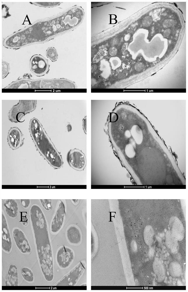
[0029] Embodiment 1: TEM sample preparation method 1

[0030] 1. Double fixation with glutaraldehyde and starvation acid: fix the spores of Fusarium oxysporum with 2.5% glutaraldehyde fixative in vacuum at 4°C for 24 hours; rinse with 0.1M sodium phosphate buffer (pH 7.2, 4°C) 6 times, 15 minutes each time for the first 4 times, 30 minutes each time for the last 2 times; then fix with 1% starvation acid solution with a mass fraction of 1% prepared by 0.1M sodium phosphate buffer (pH 7.2) for 4 hours at 4°C; M sodium phosphate buffer (pH 7.2, 4°C) was rinsed 6 times, the first 4 times were 15 minutes each time, and the last 2 times were 30 minutes each time.

[0031] 2. Pre-staining: place the fixed Fusarium oxysporum spores in 0.5% uranyl acetate aqueous solution at 4°C overnight (12 hours); rinse with 0.1M sodium phosphate buffer (pH 7.2, 4°C) for 6 15 minutes each time for the first 4 times, and 30 minutes each time for the last 2 times.

[0032] 3. Dehydration: dehydrate ...

Embodiment 2

[0035] Embodiment 2: TEM sample preparation method 2

[0036] 1. Double fixation with glutaraldehyde and starvation acid: fix the spores of Fusarium oxysporum with 2.5% glutaraldehyde fixative in vacuum at 4°C for 24 hours; rinse with 0.1M sodium phosphate buffer (pH 7.2, 4°C) 6 times, 15 minutes each time for the first 4 times, 30 minutes each time for the last 2 times; then fix with 1% starvation acid solution with a mass fraction of 1% prepared by 0.1M sodium phosphate buffer (pH 7.2) for 4 hours at 4°C; M sodium phosphate buffer (pH 7.2, 4°C) was rinsed 6 times, the first 4 times were 15 minutes each time, and the last 2 times were 30 minutes each time.

[0037] 2. Dehydration and pre-staining: dehydrate the fixed Fusarium oxysporum spores with 30% ethanol aqueous solution at 4°C for 20 minutes, 50% ethanol aqueous solution at 4°C for 20 minutes, and 70% ethanol solution. The mass fraction 0.5% uranyl acetate solution prepared in aqueous solution was pre-stained at 4°C ov...


PUM

PropertyMeasurementUnit
quality scoreaaaaaaaaaa

Abstract

The invention discloses a preparation method of a transmission electron microscope sample. According to the characteristic that fusarium oxysporum spores are thick, a glutaraldehyde and osmic acid double-fixation method is adopted for fixation, and a continuous and uniform spore dispersion technology is matched, so that fixation is more sufficient. After a glutaraldehyde and osmic acid double-fixation method is adopted for fixation, an overnight pre-staining method is added, so that the structure of each organelle in a spore cell is highlighted. Compared with the prior art, the method has the characteristics of clear imaging and obvious contrast of organelles (especially mitochondria) in fusarium oxysporum spores. Therefore, technical support is provided for further research on internal structure change of the fusarium oxysporum spores, and a microscopic level basis is provided for subsequent adoption of a targeted fusarium oxysporum prevention and control strategy.

Description

technical field [0001] The invention belongs to the technical field of transmission electron microscopy, and in particular relates to a preparation method of a transmission electron microscopy sample. Background technique [0002] Fusarium oxysporum is the main cause of banana wilt. Among them, race 1 and race 4 seriously threaten global banana production (Siddhesh, International Journal of Pest Management, 2015, 61(3):250-263). Its soil-borne characteristics make it very difficult to control or even eliminate Fusarium oxysporum (Carvalhais, Frontier in plant science, 2019, 10). As the disease progresses, the leaves of the plant begin to turn yellow and gradually wither. As the disease progresses further, the leaves will drop and form a ring around the pseudostem. Marginal ruffling appears on young leaves (Berg, Molecular Plant Pathology, 2010, 8(3)). Once banana plants are infected with Fusarium oxysporum, there are currently no effective control measures. This difficu...

Claims


Application Information

Patent Timeline
Patent Type & Authority Applications(China)
IPC IPC(8): G01N23/04G01N1/30G01N1/28
CPCG01N23/04G01N1/30G01N1/28G01N2001/302G01N2001/305
Inventor 李建雄邓汝芳田志宏朱志炎贾永霞
Owner SOUTH CHINA BOTANICAL GARDEN CHINESE ACADEMY OF SCI
Learn More