Multi-mode X-ray imaging equipment for detecting foreign matters in soft tissues and method thereof

An imaging device and X-ray technology, applied in the field of biomedicine, can solve the problems of low spatial resolution of point projection images (0.2-0.5mm, unfavorable early detection and diagnosis and treatment of diseases, and inability to clearly reflect the characteristics of lesions, etc., so as to reduce medical expenses. and national medical insurance expenditures, reducing testing time and costs, and timely and accurate judgment and treatment effects

Pending Publication Date: 2021-07-27
THE SECOND PEOPLES HOSPITAL OF SHENZHEN
View PDF7 Cites 0 Cited by
  • Summary
  • Abstract
  • Description
  • Claims
  • Application Information

AI Technical Summary

Problems solved by technology

For example, breast cancer, breast fibroadenoma, mammary gland hyperplasia, articular cartilage degeneration, and arthritis are all very difficult to diagnose and differentiate
[0005] Secondly, the existing X-ray imaging device is limited by the millimeter focal spot ray source and the electronic recording device (such as flat panel detector, scintillator array, etc.) (0.2-0.5mm), which cannot clearly reflect the appearance of some small-scale but highly indicative lesion features (such as calcifications in the breast) or the morphology of tissues and organs with porous structures (such as lungs, bones), which is not conducive to Early detection, diagnosis and treatment of related diseases (such as breast cancer, emphysema, etc.)

Method used


Image

  • Multi-mode X-ray imaging equipment for detecting foreign matters in soft tissues and method thereof
  • Multi-mode X-ray imaging equipment for detecting foreign matters in soft tissues and method thereof
  • Multi-mode X-ray imaging equipment for detecting foreign matters in soft tissues and method thereof

Examples


Embodiment 1

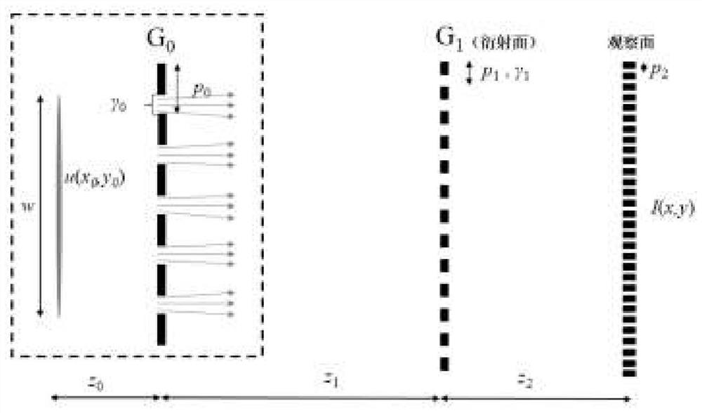
[0060] Such as figure 1 and figure 2 As shown, the multi-modal X-ray imaging device for detecting foreign bodies in soft tissues utilizes three gratings of the pfx-X-ray detector to realize clinical X-ray imaging.

[0061] Multimodal X-ray imaging devices used to detect soft tissue foreign bodies include:

[0062] A light source that produces X-rays;

[0063] Using an X-ray absorption grating to separate the X-rays incident from the light source into a source grating of a plurality of line light sources;

[0064] A phase grating capable of producing a phase distribution of the X-ray wavefront to produce regular vertical stripes downstream of the grating;

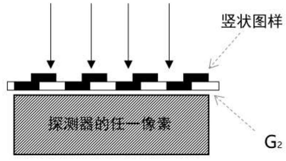
[0065] Adopt X-ray absorption grating, equipped with high-precision step displacement controller, convert the attenuation, distortion, or fuzzy information of the fringe pattern into an analysis grating of downstream X-ray intensity information;

[0066] An X-ray detector that receives X-rays and converts them into digi...


PUM


Abstract

The invention relates to the biomedical field and medical equipment technology, and discloses multi-mode X-ray imaging equipment and method for detecting foreign matters in soft tissues. The imaging detection method comprises the following steps: a working state adjustment step, a sample collection step, a signal analysis step, an image enhancement step and the like. The imaging equipment comprises a working state adjusting system, a data acquisition system, a computer data processing system and the like. According to the method, information of lesion in three aspects including the absorption capacity of a sample, electron density distribution and small-angle scattering capacity can be obtained, and by integrating and calculating the information of the absorption capacity and the small-angle scattering capacity of the sample, an R value image with certain single-angle substance identification capacity can be obtained; and therefore, the detection of common foreign matters is more comprehensive. In different subcutaneous levels, different comparison results have different advantages on identification of different types of foreign matters.

Description

technical field [0001] The invention relates to the field of biomedicine and medical equipment technology, in particular to a multi-mode X-ray imaging device and a method thereof for detecting foreign bodies in soft tissues. Background technique [0002] X-ray transmission imaging and its related X-ray computed tomography (CT) technology, as a basic method for obtaining medical images, has low cost, With the characteristics of fast speed and high efficiency, it is widely used in biomedical research and disease diagnosis and treatment. [0003] At present, the X-ray imaging devices commonly used in hospitals are based on the principle of point projection imaging, using the difference in X-ray absorption ability caused by the element composition and density changes inside the detected object to obtain images with a certain contrast (or contrast). Although the absorption contrast images given by the existing X-ray imaging devices contain rich information about the internal str...

Claims


Application Information

Patent Timeline
Patent Type & Authority Applications(China)
IPC IPC(8): A61B6/12A61B6/00
CPCA61B6/12A61B6/52A61B6/5235
Inventor 尹美芳李晶邓锴袁鸣洲骆永全张大勇吴军
Owner THE SECOND PEOPLES HOSPITAL OF SHENZHEN
Why Eureka
Try Eureka