Systems and methods for reducing colored noise in medical images using deep neural network

A deep neural network and medical image technology, applied in the field of deep neural network to reduce noise in medical images, can solve problems such as reducing image clarity and resolution, noise reduction, and affecting diagnostic quality

Pending Publication Date: 2021-09-03
GE PRECISION HEALTHCARE LLC
View PDF0 Cites 1 Cited by
  • Summary
  • Abstract
  • Description
  • Claims
  • Application Information

AI Technical Summary

Problems solved by technology

[0003] Medical images obtained with certain imaging modalities, such as MRI, can contain one or more types of noise, which can reduce image clarity and resolution
Presence of noise in medical images can affect diagnostic quality
Specifically, k-space sampling patterns, image reconstruction, and postprocessing can produce medical images with colored noise (e.g., noise that is not uniformly

Method used


Image

  • Systems and methods for reducing colored noise in medical images using deep neural network
  • Systems and methods for reducing colored noise in medical images using deep neural network
  • Systems and methods for reducing colored noise in medical images using deep neural network

Examples


Embodiment Construction

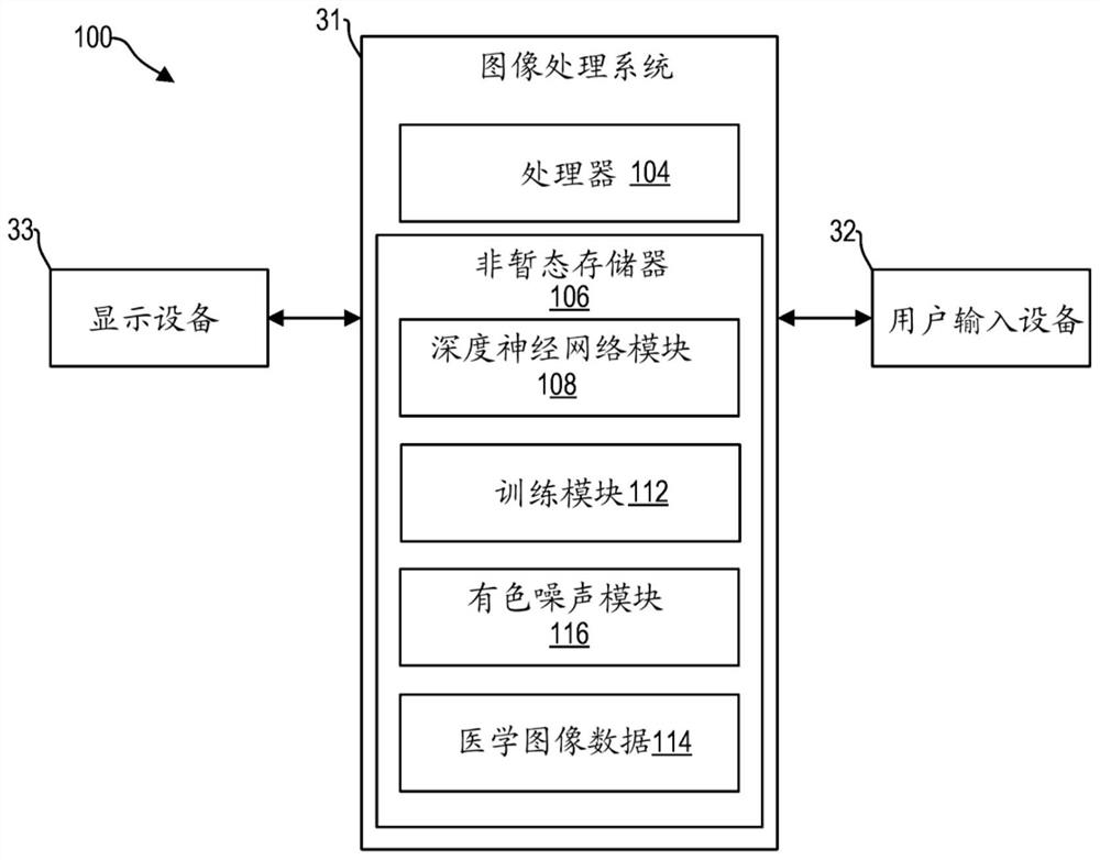
[0018] In magnetic resonance imaging (MRI), a subject is placed in a magnet. The subject is a human (alive or dead), an animal (alive or dead), or a human or part of an animal. When a subject is in a magnetic field generated by a magnet, the magnetic moments of nuclei such as protons try to align with the field, but precess around the field in random order at the Larmor frequency of the nuclei. The magnetic field of the magnet is referred to as B0 and extends in the longitudinal or z direction. During the acquisition of MRI images, a magnetic field in the x-y plane and close to the Larmor frequency (called the excitation field B1) is generated by a radio frequency (RF) coil and can be used to rotate the net magnetic moment Mz of the nucleus from the z direction or "Tilt" to the transverse or x-y plane. After the excitation signal B1 is terminated, the nucleus emits a signal called the MR signal. To generate images of a subject using MR signals, magnetic field gradient pulse...


PUM


Abstract

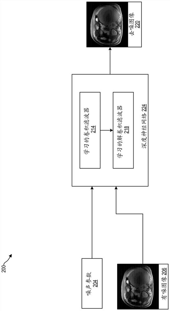
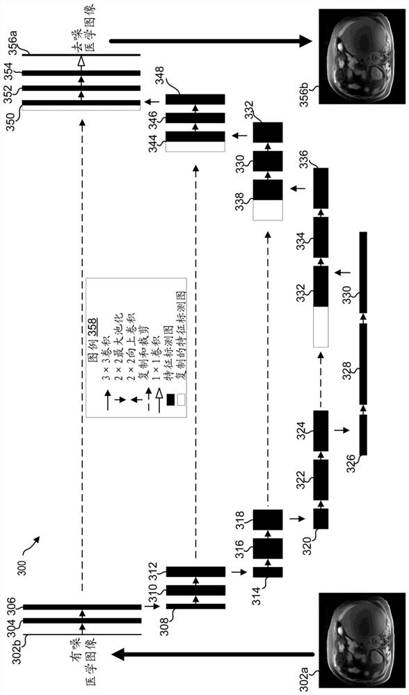
Methods and systems are provided for de-noising medical images using deep neural networks. In one embodiment, a method comprises receiving a medical image acquired by an imaging system, the medical image comprising colored noise; mapping the medical image to a de-noised medical image using a trained convolutional neural network (CNN); and displaying the de-noised medical image via a display device. The deep neural network may thereby reduce colored noise in the acquired noisy medical image, increasing a clarity and diagnostic quality of the image.

Description

technical field [0001] Embodiments of the subject matter disclosed herein relate to processing medical images, such as magnetic resonance images (MRI), CT images, etc., and more particularly to reducing noise in medical images using deep neural networks. Background technique [0002] Medical imaging systems are commonly used to obtain anatomical and / or internal physiological information of a subject, such as a patient. For example, medical imaging systems may be used to obtain medical images of a patient's skeletal structure, brain, heart, lungs, and various other features. A medical image may be an image generated by a medical imaging system. Medical imaging systems may include magnetic resonance imaging (MRI) systems, computed tomography (CT) systems, x-ray systems, ultrasound systems, and various other imaging modalities. [0003] Medical images obtained by certain imaging modalities, such as MRI, may contain one or more types of noise, which can reduce image clarity an...

Claims


Application Information

Patent Timeline
IPC IPC(8): G06T5/00G06N3/04G06N3/08
CPCG06T5/002G06N3/084G06T2207/20081G06T2207/20084G06T2207/10088G06T2207/30016G06N3/045A61B6/52A61B8/5215G06T2207/20056G06T2207/30004G06T7/0012A61B6/5258G06T2207/10024G06T2207/20182
Inventor 丹尼尔·利特威勒王新增阿里·埃尔索兹罗伯特·马克·莱贝尔埃尔辛·拜拉姆格雷姆·科林·麦金农
Owner GE PRECISION HEALTHCARE LLC
Why Eureka
Try Eureka