
Method for separating inner ear FZD10 positive glial cells

A glial cell, positive technology, applied in the field of cell separation and purification, can solve the problems of imperfection, prolonged experiment time, cumbersome process, etc., to achieve the effect of easy operation, ensure cell activity, and avoid serious damage

Pending Publication Date: 2021-09-17
山东省第二人民医院(山东省耳鼻喉医院、山东省耳鼻喉研究所)
View PDF3 Cites 0 Cited by
  • Summary
  • Abstract
  • Description
  • Claims
  • Application Information

AI Technical Summary

Problems solved by technology

[0004] However, due to the complex anatomical structure of the cochlea and the limitations of research techniques, there is still a lack of methods for the isolation and purification of cochlear glial cells.
The first step in the isolation and purification of glial cells is the digestion and separation of cochlear modioli. In most previous literatures, the introduction of the digestion method of cochlear modioli in the inner ear is not perfect, such as the type, concentration, and digestion of enzymes used for digestion. Conditions, cell pipetting methods, etc. are not described in detail (Petitpre et al., 2018)
Sun et al. used a two-step digestion method in the digestion process of the snail shaft, and the digestion time of each step was as long as 30 minutes, and there were many kinds of reagents used, and the process was cumbersome
[0005] In addition, in terms of purification of glial cells, previous studies could only isolate whole modioli cells and study their differentiation into neurons
However, the cells in the modiolus include neurons, glial cells, fibroblasts and other cell types, which makes it difficult to study the proliferation and differentiation characteristics of glial cells
Some researchers take advantage of the characteristics that neurons cannot proliferate and pass on, and remove the neurons in the isolated cochlear modiolus cells by first cultured into spheres for more than three generations. Greatly prolong the experiment time and reduce the activity of glial cells to a certain extent
In addition, since fibroblasts in the inner ear also have the ability to proliferate, this method does not remove modioli fibroblasts very well (McLean, McLean, Eatock, & Edge, 2016)

Method used


Image

  • Method for separating inner ear FZD10 positive glial cells
  • Method for separating inner ear FZD10 positive glial cells

Examples


Embodiment 1

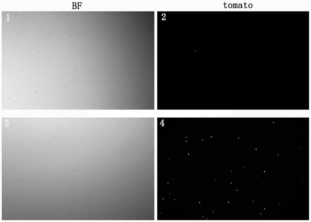
[0079] (1) Take 20 FZD10-creER+ / - / Rosa26R-tdTomato+ / - double-positive suckling mice 3 days after birth, spray them with alcohol and put them in a biological safety cabinet for operation. Remove the heads of the suckling mice and split them sagittally from the middle of the skull Open, remove the brain parenchyma, cut off the bilateral temporal bones where the cochlea is located, quickly put it into a petri dish filled with cold Hanks solution, carefully dissect the cochlea under a dissecting microscope, remove the cochlea to expose the internal structure of the cochlea, and carefully tear off the periphery The spiral ligament was removed and the organ of Corti was removed, and the remaining modiolus was transferred to a 15 ml centrifuge tube containing cold Hanks solution. Put the 15mL centrifuge tube containing the worm shaft tissue into the centrifuge and centrifuge at 1500rpm for 5 minutes.

[0080] (2) Prepare the enzymatic hydrolysis solution required for digesting cochle...

Embodiment 2

[0092] (1) Take 20 FZD10-creER+ / - / Rosa26R-tdTomato+ / - double-positive suckling mice two days after birth, spray them with alcohol and put them in a biological safety cabinet for operation. The heads of the suckling mice are removed and split sagittal from the middle of the skull , remove the brain parenchyma, cut off the bilateral temporal bones where the cochlea is located, quickly put it into a petri dish filled with cold Hanks solution, carefully dissect the cochlea under a dissecting microscope, remove the cochlea shell to expose the internal structure of the cochlea, and carefully tear off the peripheral The ligament was spiraled and the organ of Corti removed, and the remaining modiolus was transferred to a 15 ml centrifuge tube containing cold Hanks solution. Put the 15mL centrifuge tube containing the worm shaft tissue into the centrifuge and centrifuge at 1500rpm for 5 minutes.

[0093] (2) Prepare the enzymatic hydrolysis solution required for digesting cochlea mandr...

Embodiment 3

[0105] (1) Take 20 FZD10-creER+ / - / Rosa26R-tdTomato+ / - double-positive suckling mice three days after birth, spray them with alcohol and put them in a biological safety cabinet for operation. The heads of the suckling mice are removed and split sagittal from the middle of the skull , remove the brain parenchyma, cut off the bilateral temporal bones where the cochlea is located, quickly put it into a petri dish filled with cold Hanks solution, carefully dissect the cochlea under a dissecting microscope, remove the cochlea shell to expose the internal structure of the cochlea, and carefully tear off the peripheral The ligament was spiraled and the organ of Corti removed, and the remaining modiolus was transferred to a 15 ml centrifuge tube containing cold Hanks solution. Put the 15mL centrifuge tube containing the worm shaft tissue into the centrifuge and centrifuge at 1500rpm for 5 minutes.

[0106] (2) Prepare the enzymatic hydrolysis solution required for digesting cochlea man...


PUM


Abstract

The invention relates to the field of cell separation and purification, in particular to a method for separating inner ear FZD10 positive glial cells. According to the method for separating the inner ear FZD10 positive glial cells, the inner ear FZD10 positive glial cells are subjected to fluorescence labeling by virtue of transgenic mouse hybridization, so that purified inner ear glial cells can be conveniently obtained subsequently by virtue of flow sorting. According to a separation and purification technology adopted by the method, the glial cells can be obtained to the maximum extent, the activity of the cells can be kept, the purity of the separated FZD10 positive glial cells reaches 98% or above, the requirements of follow-up experiments can be met, analysis and purification process is simple and rapid, and the method is very suitable for follow-up experiment research.

Description

technical field [0001] The invention relates to the field of cell separation and purification, in particular to a method for isolating inner ear Frizzled10 (FZD10) positive glial cells. Background technique [0002] Deafness is a major global public health problem. About 500 million people around the world suffer from disabling hearing loss, accounting for 6.5% of the world's total population, and there is an upward trend. Among them, patients with sensorineural deafness accounted for about 65%. Sensorineural deafness refers to the location of the lesion in the cochlea and the auditory pathway behind it, which is mainly caused by the degeneration of cochlear hair cells and spiral neurons. Unlike conductive deafness caused by external auditory canal or middle ear lesions, a considerable part of sensorineural deafness is often permanent and difficult to reverse. The current treatment for sensorineural hearing loss mainly adopts cochlear implantation. Cochlear implants play ...

Claims


Application Information

Patent Timeline
IPC IPC(8): C12N5/079
CPCC12N5/0622C12N2509/00C12N2509/10C12N2501/115C12N2501/105C12N2501/11C12N2501/91
Inventor 刘闻闻王嫚徐磊王海波
Owner 山东省第二人民医院(山东省耳鼻喉医院、山东省耳鼻喉研究所)
Learn More