Endogastric biopsy histopathologic imaging method based on stimulated Raman scattering

A technology of stimulated Raman scattering and imaging methods, applied in Raman scattering, image enhancement, image analysis, etc., can solve the problems of time lag, labor-intensive processing, complicated sample pretreatment, etc., and achieve the effect of fast imaging speed

Active Publication Date: 2021-09-24
FUDAN UNIV
View PDF3 Cites 2 Cited by
  • Summary
  • Abstract
  • Description
  • Claims
  • Application Information

AI Technical Summary

Problems solved by technology

[0005] The purpose of the present invention is to provide a gastric endoscopic biopsy histopathological imaging method with fast imaging speed and high

Method used


Image

  • Endogastric biopsy histopathologic imaging method based on stimulated Raman scattering
  • Endogastric biopsy histopathologic imaging method based on stimulated Raman scattering
  • Endogastric biopsy histopathologic imaging method based on stimulated Raman scattering

Examples


Embodiment 1

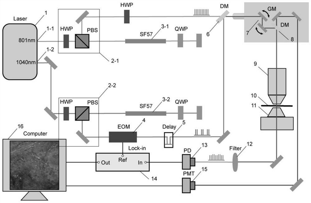
[0028] Build a stimulated Raman scattering microscopy imaging system, such as figure 1 shown. In the system, the laser 1 generates femtosecond pulsed laser, and the pump light output port 1-1 can be femtosecond laser with a tunable wavelength of 680nm-1300nm as the pump light, and the Stokes light output port 1-2 at the other end outputs Femtosecond laser with a fixed wavelength of 1040nm, as Stokes light. After the pump light and Stokes light are adjusted through the combination 2-1 and 2-2 of the half-wave plate and the polarization beam splitter prism, the linear chirp process is completed through the SF57 dispersion glass 3-1 and 3-2, so that the flying The second light is stretched into picosecond light, and the spectrum is arranged in time and space during the chirping process, providing a full width at half maximum of 15cm for the stimulated Raman scattering system -1spectral resolution. Then, the Stokes light is digitally modulated by the electro-optic modulator 4 a...

Embodiment 2

[0032] In this embodiment, lipid, protein and collagen are selected as the substances to be detected. With reference to Example 1, the rapid histopathological imaging method of gastric biopsy tissue includes the following steps:

[0033] S1. Obtain the experimental parameters of the respective Raman peaks in the stimulated Raman scattering microscope system from the actual situation of imaging of lipids and proteins. The parameters include: the fixed Stokes wavelength is 1040nm, and the pump wavelength is Choose 801nm, and the wave number corresponding to the corresponding time delay is selected as 2845cm -1 and 2930cm -1 , the specific delay position is obtained according to the actual experimental conditions;

[0034] S2. First set up the two outputs of the laser, and then set the corresponding wave number of the relative time delay between Stokes light and pump light to 2845cm -1 , after selecting the focal plane, move the sample translation stage 10 to find the desired ...


PUM


Abstract

The invention belongs to the technical field of nonlinear optical imaging, and particularly relates to an endogastric biopsy histopathology imaging method based on stimulated Raman scattering. According to the method, the histopathologic image information of stomach biopsy can be obtained in a short time by utilizing the characteristics of rapidness, no processing and no marking of stimulated Raman scattering microimaging. According to the invention, the stimulated Raman scattering microscopy technology is clinically applied to gastroscope endoscopic biopsy for the first time, and compared with the existing traditional histopathology technology, the method has the advantages that the imaging speed is high, the imaging quality is high, pretreatment is not needed, original tissues are noninvasively reserved, and each plane can be imaged within a certain depth.

Description

technical field [0001] The invention belongs to the technical field of nonlinear optical imaging, in particular to a gastric endoscopic biopsy histopathological imaging method based on stimulated Raman scattering. Background technique [0002] Gastric cancer is a major disease affecting human health. According to the latest annual report of the National Cancer Registry Center, the number of new cases of gastric cancer exceeds 680,000 each year, and about 500,000 people die, making it the second deadliest tumor in my country. The key to the prevention and control of gastric cancer is early diagnosis and early treatment. Timely detection of early cancer, delay and reversal of precancerous lesions can effectively reduce the incidence and mortality of gastric cancer. The occurrence of gastric cancer goes through multiple steps such as chronic inflammation-precancerous lesions-early cancer-invasive cancer. The development of gastric cancer usually requires decades of developme...

Claims


Application Information

Patent Timeline
IPC IPC(8): G01N21/65G06T3/40G06T7/00
CPCG01N21/65G06T7/0012G06T3/4038G06T3/4053G01N2021/655G06T2207/10024G06T2207/30004
Inventor 季敏标刘至杰胡皓敖建鹏周平红
Owner FUDAN UNIV
Why Eureka
Try Eureka