Immunohistochemical cell image cell nucleus segmentation and counting method and system

A technology of immunohistochemistry and counting methods, which is applied in image analysis, image data processing, image enhancement, etc., can solve the problems of deep learning algorithm, such as large amount of calculation, difficulty in parameter selection, difficulty in obtaining labeled samples of immunohistochemical cell images, etc.

Active Publication Date: 2021-10-01
北京透彻未来科技有限公司
View PDF4 Cites 5 Cited by
  • Summary
  • Abstract
  • Description
  • Claims
  • Application Information

AI Technical Summary

Problems solved by technology

Nucleus detection and segmentation is the key first step in medical image analysis. However, due to the existence of uneven staining, irregular cell shape, adhesion between nuclei, and impurity noise, accurate cell nucleus segmentation and counting still face many challenges. It is a big challenge. The existing cell image segmentation technology mainly adopts deep learning algorithm, which requires a large amount of training data. However, it is extremely difficult to obtain labeled samples of immunohistochemical cell images, and the training data is insufficient, resulting in insufficient accuracy.
At the same time, the deep learning algorithm has a large amount of calculation and requires high computer resources.
However, the segmentation method based on non-deep learning algorithm needs to regulate a large number of parameters, and the selection of parameters is more difficult.

Method used


Image

  • Immunohistochemical cell image cell nucleus segmentation and counting method and system
  • Immunohistochemical cell image cell nucleus segmentation and counting method and system
  • Immunohistochemical cell image cell nucleus segmentation and counting method and system

Examples


Embodiment 1

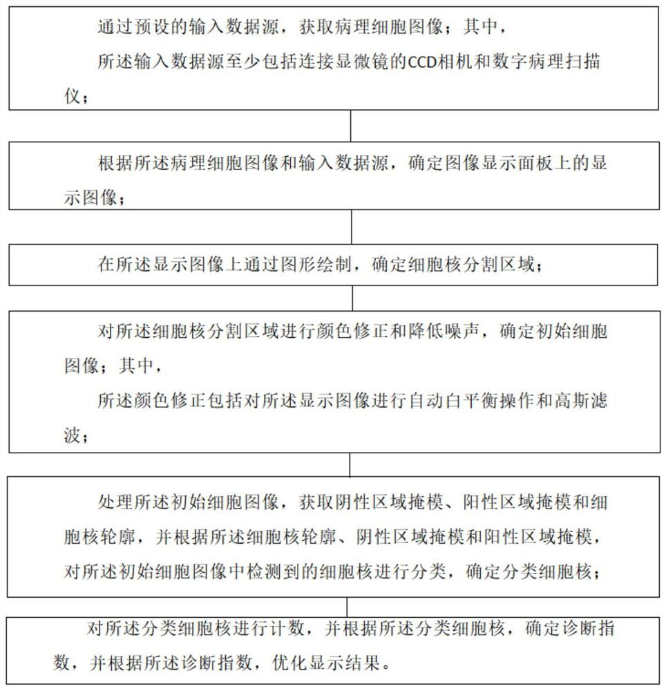
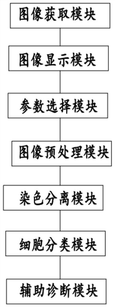
[0104] according to Figure 1-9 As shown, the present invention provides a method for segmenting and counting cell nuclei in immunohistochemical cell images, which is characterized in that it includes:

[0105] Obtain pathological cell images through preset input data sources; where,

[0106] The input data source includes at least a CCD camera connected to a microscope and a digital pathology scanner;

[0107] Determine the display image on the image display panel according to the pathological cell image and the input data source;

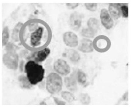
[0108] Determining the segmented area of ​​the cell nucleus by graphically drawing on the displayed image;

[0109] Carrying out color correction and noise reduction on the cell nucleus segmentation area to determine the initial cell image; wherein,

[0110] The color correction includes performing automatic white balance operation and Gaussian filtering on the displayed image;

[0111] Processing the initial cell image to obtain a negative ar...

Embodiment 2

[0116] according to Figure 1-9 As shown, the technical solution provides an embodiment, the determination of the display image on the image display panel according to the pathological cell image and the input data source further includes:

[0117] When the input data source is a CCD camera connected to a microscope, the pathological cell image is updated in real time based on the preset CCD camera sampling, and the display image is determined;

[0118] When the input data source is a digital pathology scanner, based on the user's needs, perform area selection on the cell image to determine the display image; wherein,

[0119] The region selection includes at least zooming and panning.

[0120] The working principle and beneficial effects of the above-mentioned technical scheme are:

[0121] The input data source of this technical solution can be a CCD camera connected to a microscope, or a digital pathological image obtained by a digital pathology scanner. In the CCD camera...

Embodiment 3

[0123] according to Figure 1-9 As shown, the technical solution provides an embodiment,

[0124] The step of determining the cell nucleus segmented area by graphically drawing on the displayed image includes:

[0125] Determining the area of ​​the cell nucleus based on the image display panel;

[0126] According to the area of ​​the nucleus, a graphic frame is drawn for the nucleus to determine the division area of ​​the nucleus; wherein,

[0127] The graphic frame includes a rectangle, a circle or an arbitrary curve.

[0128] In actual implementation, the present invention also adopts the display area segmented according to the graphic frame and the cell to determine the segmentation parameters; wherein,

[0129] The segmentation parameters include circular parameters and rectangular parameters; wherein,

[0130] The circle parameter is used to set the minimum area of ​​the nucleus and the color threshold of the positive level;

[0131] The rectangle parameter is used t...


PUM


Abstract

The invention provides an immunohistochemical cell image cell nucleus segmentation and counting method, which comprises the following steps: acquiring a pathological cell image through a preset input data source; determining a display image on an image display panel according to the pathological cell image and an input data source; determining a cell nucleus segmentation area on the display image through graph drawing; performing color correction and noise reduction on the cell nucleus segmentation region, and determining an initial cell image; processing the initial cell image, obtaining a negative area mask, a positive area mask and a cell nucleus contour, classifying cell nucleuses detected in the initial cell image according to the cell nucleus contour, the negative area mask and the positive area mask, and determining classified cell nucleuses; and counting the classified cell nucleuses, determining a diagnosis index according to the classified cell nucleuses, and optimizing a display result according to the diagnosis index.

Description

technical field [0001] The present invention relates to the technical field of cell nucleus segmentation and counting methods and systems for immunohistochemical cell images, in particular to a cell nucleus segmentation and counting method and system for immunohistochemical cell images. Background technique [0002] At present, pathological diagnosis is the most reliable method of tumor examination and is called the "gold standard". Cancerous nuclei are significantly different from normal nuclei in size and shape, and can be visualized by specific staining. Currently, pathological diagnosis is mostly done through observation under a microscope, which requires very high expertise, takes a lot of time, and produces subjective differences. Computer-aided diagnosis can provide high-speed, repeatable medical image analysis, and the results are more objective and accurate. Nucleus detection and segmentation is a critical first step in medical image analysis. However, due to the ...

Claims


Application Information

Patent Timeline
Patent Type & Authority Applications(China)
IPC IPC(8): G06T7/11G06T7/13G06T7/136G06T5/00G06T7/62
CPCG06T7/11G06T7/13G06T7/136G06T5/002G06T5/009G06T7/62G06T2207/10061G06T2207/30024G06T2207/30242
Inventor 王书浩王朗
Owner 北京透彻未来科技有限公司
Why Eureka
Try Eureka