Confocal endoscope imaging device

An imaging device and endoscope technology, which is applied in the field of confocal imaging, can solve the problems of poor deep tissue imaging effect, small imaging depth of endoscope, and inability to use internal tissues, achieve high resolution, and overcome the generally poor imaging depth. Small, easily adjustable effect

Pending Publication Date: 2021-10-12
济南国科医工科技发展有限公司
View PDF1 Cites 0 Cited by
  • Summary
  • Abstract
  • Description
  • Claims
  • Application Information

AI Technical Summary

Problems solved by technology

However, it cannot be used for the detection of internal tissues, such as the digestive tract
[0004] At present, endoscopes are usually used clinically to detect internal tissues such as digestive tract tumors, but the imaging depth of endoscopes is generally small, and the imaging effect on deep tissues is poor
[0005] Therefore, using confocal imaging has the characteristics of high resolution and the ability to achieve imaging effects in deep tissues. Applying confocal imaging to endoscopic imaging will be of great significance for the detection of internal tissues of the human body, such as the detection of gastrointestinal lesions, but there is currently a lack of reliable plan

Method used


Image

  • Confocal endoscope imaging device
  • Confocal endoscope imaging device
  • Confocal endoscope imaging device

Examples


Embodiment 1

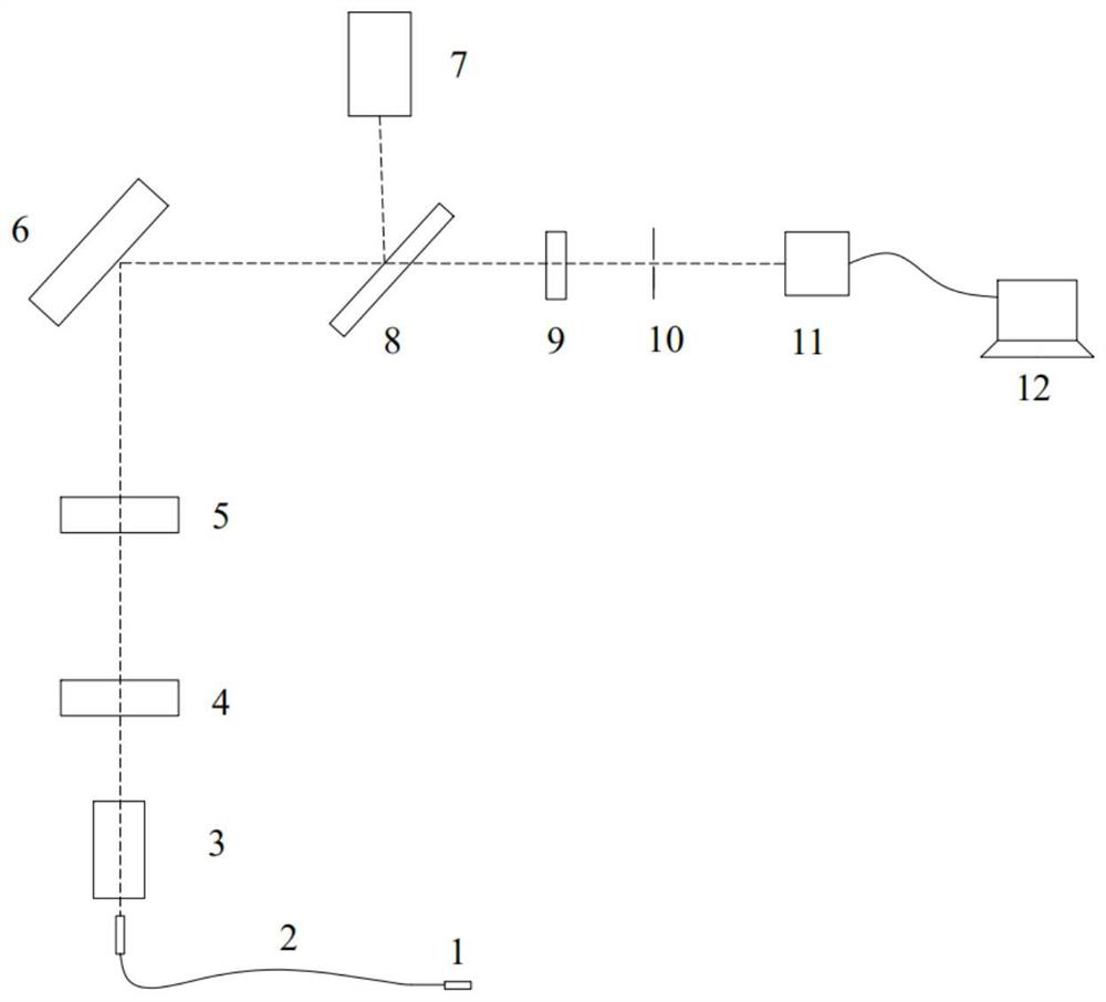
[0048] refer to Figure 1-Figure 3 , a confocal endoscopic imaging device of this embodiment, comprising:

[0049] Endoscopic objective lens 31, endoscopic optical fiber bundle 2, coupling objective lens 3, first lens 4, second lens 5, high-speed scanning galvanometer 6, laser 7, dichroic mirror 8, third lens 9, pinhole 10, photoelectric Multiplier tube 11 and computer 12

[0050]The laser light emitted by the laser 7 is reflected by the dichroic mirror 8 to the high-speed scanning galvanometer 6, then passes through the second lens 5, the first lens 4, and the coupling objective lens 3 in turn, and then is coupled with the first end of the endoscopic optical fiber bundle 2, and then passed through The endoscopic objective lens 31 at the second end of the endoscopic optical fiber bundle 2 irradiates the tissue; the reflected light of the tissue returns to the dichroic mirror 8 through the original path, and the reflected light passes through the third lens 9 and the pinhole a...

Embodiment 2

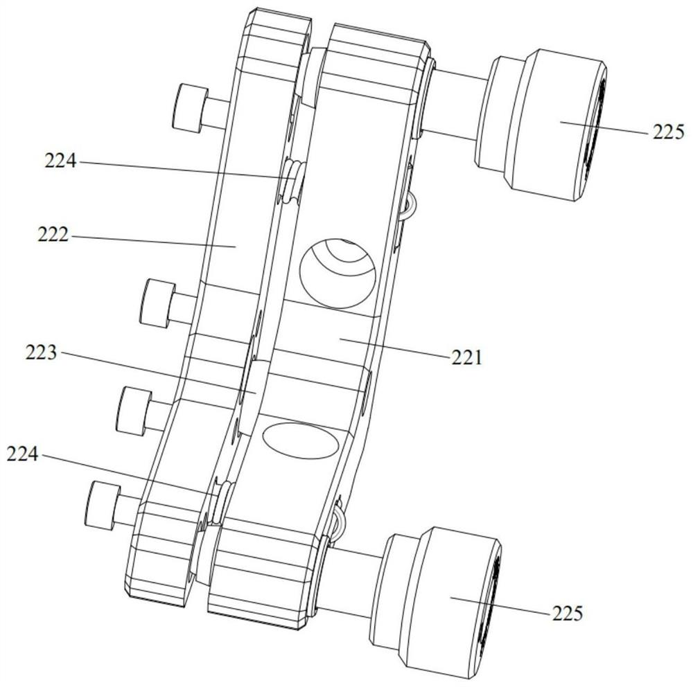
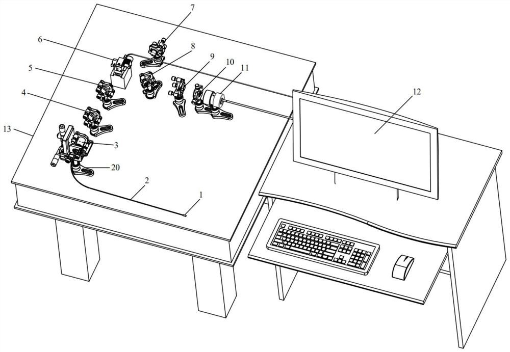
[0056] As a further improvement on the basis of Example 1, refer to Figure 4-Figure 12 , in this embodiment, the confocal endoscopic imaging device also includes an optical table 13 and an optical fiber bundle adjustment frame 20 arranged on the optical table 13, and the first end of the endoscopic optical fiber bundle 2 is arranged on the optical fiber bundle adjustment frame 20 . The optical fiber bundle adjustment frame 20 is used to adjust the position of the first end of the endoscopic optical fiber bundle 2 to ensure that the endoscopic optical fiber bundle 2 is facing the coupling objective lens 3 to achieve efficient beam coupling.

[0057] Wherein, the optical fiber bundle adjustment frame 20 includes a lift adjustment bracket 21 connected to the optical platform 13, an angle adjustment bracket 22 arranged on the lift adjustment bracket 21, a mounting plate 23 arranged on the angle adjustment bracket 22, and a mounting plate 23 arranged on the mounting plate 23. An ...


PUM


Abstract

The invention discloses a confocal endoscope imaging device. The device comprises an endoscopic objective lens, an endoscopic optical fiber bundle, a coupling objective lens, a first lens, a second lens, a high-speed scanning galvanometer, a laser, a dichroscope, a third lens, a pinhole, a photomultiplier tube and a computer. According to the device, confocal imaging and endoscopic imaging are combined, the endoscopic optical fiber bundle and the endoscopic objective lens are arranged, so that the device can extend into a digestive tract of a human body for detection, the advantages of high resolution of the confocal imaging and realization of an imaging effect of deep tissues can be fully utilized, so that lesion detection of the digestive tract can be realized on a cellular level, and the defects that the imaging depth of an existing clinical endoscope is generally small, and the imaging effect on deep tissue is poor are overcome. The invention further provides an optical fiber bundle adjusting frame which can conveniently adjust the position of a first end of the endoscopic optical fiber bundle, so that the endoscopic optical fiber bundle directly faces the coupling objective lens, and light beam coupling is achieved.

Description

technical field [0001] The invention relates to the field of confocal imaging, in particular to a confocal endoscope imaging device. Background technique [0002] Digestive tract tumors are characterized by high morbidity and high lethality, but the survival rate of early digestive tract tumors can reach more than 50% after cure. [0003] The precise pinhole of the confocal imaging device filters out stray light, and only the points on the focal plane of the sample participate in the imaging. It has the characteristics of high resolution and clear images, and can be used to observe the occurrence, development, curative effect and skin lesions of skin diseases. Observation of internal organization cannot be achieved. For example, Chinese patent CN202022435855.0 discloses a reflective confocal imaging device, which adopts the principle of confocal imaging and can be used for observation of skin diseases. However, it cannot be used for the detection of internal tissues, such ...

Claims


Application Information

Patent Timeline
Patent Type & Authority Applications(China)
IPC IPC(8): A61B1/00A61B1/04A61B1/07
CPCA61B1/00188A61B1/04A61B1/07
Inventor 冷冰李旺鑫陈永超臧志刚王悦
Owner 济南国科医工科技发展有限公司
Why Eureka
Try Eureka