
In-tissue visual proximity marking method

A labeling method and tissue technology, applied in the field of protein tracking imaging, can solve the problems of highly dynamic changes in difficult inter-neuron connections, and achieve the effect of avoiding the influence of non-specific signals

Pending Publication Date: 2022-01-28
SHANGHAI TECH UNIV
View PDF10 Cites 0 Cited by
  • Summary
  • Abstract
  • Description
  • Claims
  • Application Information

AI Technical Summary

Problems solved by technology

But these methods rely on the persistence of chemical and electrical synapses, making it difficult to track the highly dynamic changes in the connections between neurons

Method used


Image

  • In-tissue visual proximity marking method
  • In-tissue visual proximity marking method
  • In-tissue visual proximity marking method

Examples


Embodiment 1

[0042] 1.1 Breeding method of mice

[0043] The experimental mice used in the present invention are ICR mouse strains. Mice were kept in SPF-grade animal facilities with 12 hours of light and 12 hours of darkness at a temperature of 22-25°C to ensure adequate supply of drinking water and food. All animal experiments were strictly complied with the rules and regulations of the Animal Care and Use Committee of ShanghaiTech University.

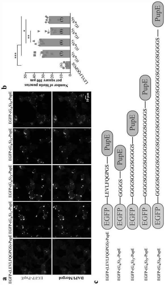
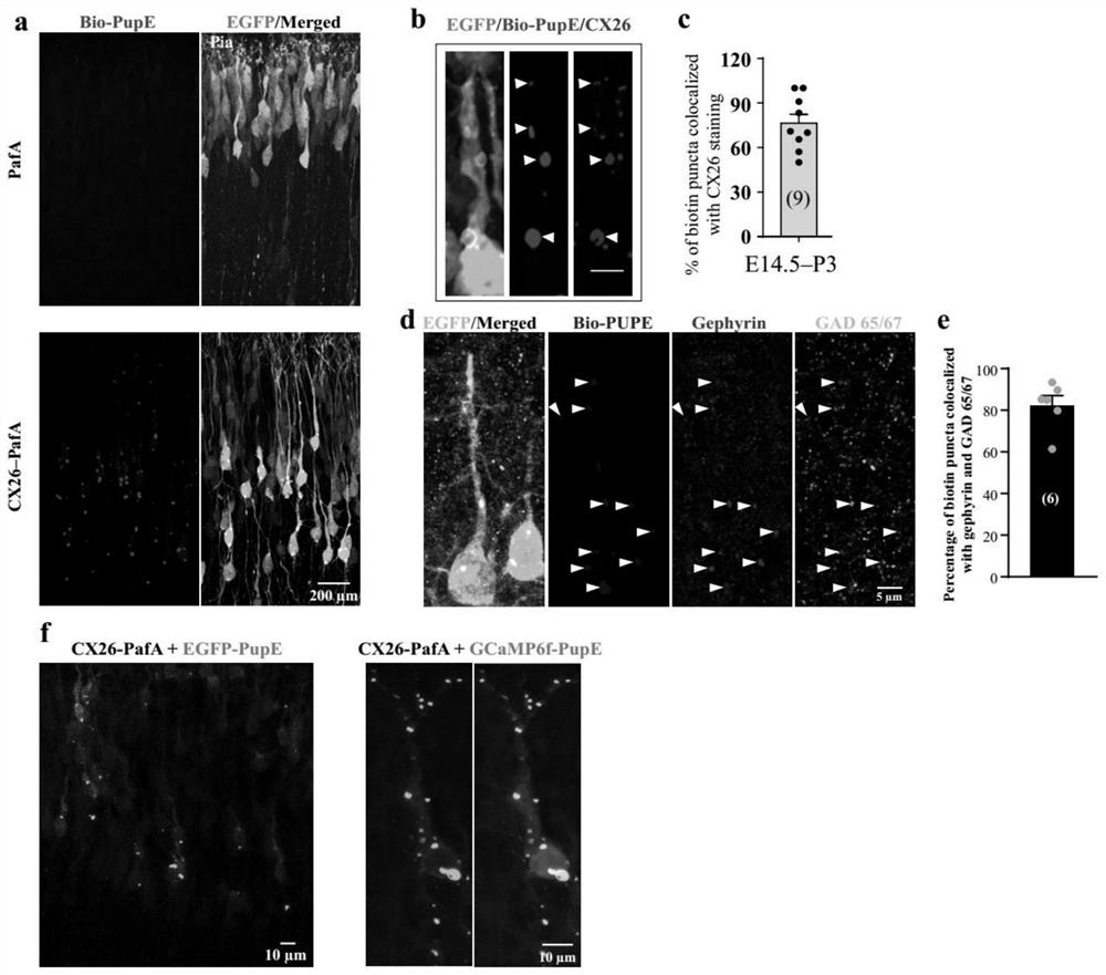
[0044] 1.2 Construction of target protein fusion PafA plasmid and fluorescent protein fusion PupE plasmid

[0045] The sequence of electrical synapse protein CX26 was amplified by PCR from the cDNA library of neonatal mouse cortex, and the sequence of inhibitory chemical synapse protein Gephyrin was cloned from the cDNA library of adult mouse cortex. Both CX26 and Gephyrin were fused to PafA using the Gly-Ser-Ser-Gly-Ser (GSSGS, as shown in SEQ ID NO: 1) sequence, and the pCAG-IRES- EGFP plasmid vector. The fluorescent protein EGFP sequence s...

Embodiment 2

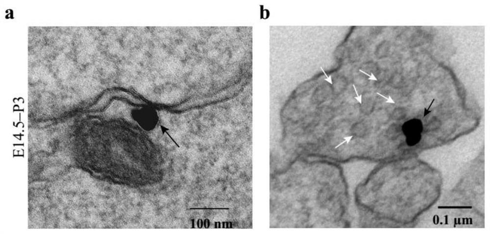
[0057] 2.1 Sample preparation for TEM

[0058] Take embryonic mice electroporated with pCAG-CX26-PafA-IRES-EGFP and pCAG-BCCP-PupE-IRES-EGFP plasmid combination, and use 2% paraformaldehyde (TED PELLA, EM grade) 2% glutaraldehyde after anesthesia Mix the solution for cardiac perfusion and quickly dissect out the mouse brain. After the brain was fixed for 3 hours, slices with a thickness of 150 μm were made and soaked in fixative solution overnight at 4°C. Brain slices with fluorescent cells were selected, soaked in 30% sucrose solution for 3 hours, and then repeatedly frozen and thawed in liquid nitrogen for 3 times. Add streptavidin (Nanogold-Streptavidin, Nanoprobes2016) with nano-gold label at a dilution ratio of 1:150, incubate at room temperature for 2 hours, and use the gold particle amplification kit (GoldEnhance EM Plus kit, Nanoprobes2114) to amplify the nano-gold label . After the immunoassay, the cortical areas in the brain slices were excised, fixed with 0.1% os...


PUM


Abstract

The invention discloses an in-tissue visual proximity marking method. The in-tissue visual proximity marker method comprises the following steps that: (1) a plasmid vector 1 and a plasmid vector 2 are transformed into a to-be-observed tissue, wherein the plasmid vector 1 carries a fusion gene for expressing a target protein and PafA, and the plasmid vector 2 carries a fusion gene for expressing a tag protein and Pup (E); and (2) after the target protein is expressed, a marking result is observed through the tag protein. The PUPIL provided by the method of the invention can be applied to the research of animal in-vivo protein visualization, and can be used for visually marking subcellular localization information of electrical synapse and inhibitory chemical synapse proteins in living tissue neurons.

Description

technical field [0001] The invention relates to the field of protein tracer imaging, in particular to a method for visualizing adjacent markers in tissues. Background technique [0002] The higher-level functions performed by the brain rely on complex networks of nerve cells that transmit signals through both chemical and electrical synapses. The labeling of chemical synapse and electrical synapse-related proteins can help us more intuitively study the function and state of synapses between neurons, and is of great significance to the study of brain development, neural circuit projections, and neurodegenerative diseases. [0003] At present, many labeling methods have been applied to the research of chemical synapses and electrical synapses, such as direct fusion of synaptic proteins with GFP (green fluorescent protein), protease BirA modification labeling, or labeling by GRASP (GFPreconstitution across synaptic partners), etc. . But these methods rely on the persistence o...

Claims


Application Information

Patent Timeline
IPC IPC(8): C12N15/85C12N15/65
CPCC12N15/85C12N15/65C12N2800/107
Inventor 谢书何水金
Owner SHANGHAI TECH UNIV
Learn More