DC cell preparation method for improving antigen presenting T cell and improving T cell killing efficiency and application thereof

A cell and antigen technology, applied in the field of preparation of DC cells, can solve the problems of low antigen presentation efficiency, unsatisfactory T cell-specific immune response effect, and inability to exert anti-tumor effect, so as to achieve high antigen presentation efficiency and improve killing effect. Efficiency, activation-promoting effect

Active Publication Date: 2022-02-11
北京荟科柘生物科技有限公司
View PDF2 Cites 0 Cited by
  • Summary
  • Abstract
  • Description
  • Claims
  • Application Information

AI Technical Summary

Problems solved by technology

At present, there are defects such as low antigen presentation efficiency and unsatisfactory T cell-spec...

Method used


Image

  • DC cell preparation method for improving antigen presenting T cell and improving T cell killing efficiency and application thereof
  • DC cell preparation method for improving antigen presenting T cell and improving T cell killing efficiency and application thereof
  • DC cell preparation method for improving antigen presenting T cell and improving T cell killing efficiency and application thereof

Examples


Embodiment 1

[0040] Example 1 Preparation method of dendritic cells that can improve antigen-presenting T cells and increase T-cell killing activity

[0041] 1. Preparation of DC cells

[0042] 1.1 Add 5ml of peripheral blood serum into a T75 bottle, and cover in the dark at 37°C for 4 hours.

[0043] 1.2 Pour fresh heparin-anticoagulated human peripheral blood into a centrifuge tube, balance, and centrifuge at 700g / min for 20 minutes (the slowest rate of decline).

[0044] 1.3 Add 1:1 D-PBS to the cell layer after centrifugation of the above-mentioned peripheral blood and mix well, slowly add the lymphocyte separation liquid along the tube wall at a ratio of 1:1 to the surface of the lymphocyte separation liquid, and keep a clear interface. After centrifugation at 800g for 15min (slow rise and fall), the centrifuge tube is divided into four layers from top to bottom.

[0045] Among them, the first layer is a D-PBS layer, the second layer is a ring-shaped milky white PBMC, the third laye...

experiment example 1

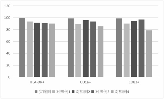
[0142] Experimental Example 1 Flow cytometric detection of DC maturation ratio

[0143] The DC cell surface markers HLA-DR+, CD1a+, CD83+ obtained in the embodiment and 4 groups of control examples were detected by flow cytometry, and the detection results were as follows: figure 1 shown. Depend on figure 1 From the results, it can be seen that the expression levels of markers on the surface of DC cells obtained in the examples were higher than those in the 4 groups of control examples. It can be concluded that the rate of obtaining mature DC in the example is higher than that in the 4 groups of control examples.

experiment example 2

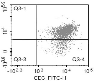
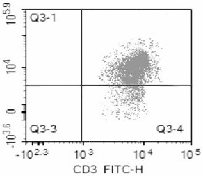
[0144] Experimental Example 2 Detection of T cells and their secretion of cytokines

[0145] 1) Use flow cytometry to detect the maturation ratio of DC-stimulated T cells obtained in the example and the 4 groups of control examples; the results are as follows Figure 2-6 shown. The results showed that the ratio of DC cells obtained in the embodiment to stimulate T cell maturation was 91.78% ( figure 2 ), the DC cells obtained in Control Example 1 stimulated T cell maturation ratio was 82.31% ( image 3 ), the ratio of DC cells obtained in Control Example 2 to stimulate T cell maturation was 74.96% ( Figure 4 ), the DC cells obtained in Control Example 3 stimulated T cell maturation ratio was 78.24% ( Figure 5 ), the ratio of DC cells obtained in Control Example 4 to stimulate T cell maturation was 62.31% ( Figure 6 ) suggest that DCs obtained by the method described in this application can more effectively activate antigen-presenting T cells.

[0146] 2) ELISA detecti...


PUM


Abstract

The invention provides a DC cell preparation method for improving antigen presenting T cells and improving T cell killing efficiency and application thereof. A tumor antigen modified by 4-phenyl isothiocyanate alpha-D-mannopyranoside glycosylation is loaded on a DC cell, SDF-1 and IL-8 chemotactic factors are added, the DC cell is chemotactic, more mature DCs are obtained, interaction between the DC and T cell is enhanced, T cell activation is further promoted, a gene expression silencing agent shRNA is introduced into the DC cell, an IDO gene is silenced, the surface antigen of the DC is obviously increased, and the proliferation reaction of T lymphocytes is enhanced and stimulated; after the DC cells and the T cells are subjected to mixed culture, the pH value is adjusted, and the DC cells and the T cells are cultured in a weak acid environment, so that the antigen presentation efficiency of the DC cells and the T cells is improved. The result shows that the antigen presentation efficiency between the DC cells and the T cells is higher, and the T cells with higher antigen specificity and higher killing efficiency can be obtained through stimulation.

Description

technical field [0001] The invention relates to the field of cell culture, in particular to a preparation method and application of a DC cell capable of strong immune regulation, enhancing antigen-presenting T cells and improving T cell killing efficiency. Background technique [0002] Dendritic cells (English: dendritic cell, DC), also known as dendritic cells, dendritic cells, are a type of white blood cells that exist in mammals. It is found in the blood and in tissues exposed to the environment, such as the skin and epithelial tissue of the nose, lungs, stomach and small intestine. They function to regulate innate and acquired immune responses to current environmental stimuli. One of its most important functions is to present antigens to T cells of the immune system after processing, so it is an antigen-presenting cell. DCs are usually distributed in a small amount in the skin and mucous membranes that are in contact with the outside world. The skin is called Langerhan...

Claims


Application Information

Patent Timeline
IPC IPC(8): C12N5/0783C12N5/0784C12N5/10C12N15/12A61K35/17A61P35/00
CPCC12N5/0639C12N5/0638C07K14/47A61K35/17A61P35/00
Inventor 朱哲张涛
Owner 北京荟科柘生物科技有限公司
Why Eureka
Try Eureka