Skin blood vessel and blood flow quantitative evaluation system and method based on optical imaging

An optical imaging and evaluation system technology, applied in the field of skin blood vessel and blood flow quantitative evaluation system, can solve problems such as one-sidedness, poor accuracy, and incomplete three-dimensional evaluation of blood vessels.

Pending Publication Date: 2022-02-15
EXPERIMENTAL RES CENT CHINA ACAD OF CHINESE MEDICAL SCI
View PDF0 Cites 0 Cited by
  • Summary
  • Abstract
  • Description
  • Claims
  • Application Information

AI Technical Summary

Problems solved by technology

[0003] At present, the observation of the morphology of dermal blood vessels and the amount of blood perfusion is still limited. On the one hand, a single method cannot evaluate the results, which is relatively one-sided and the accuracy is not good. A comprehensive evaluation system for blood flow in blood vessels has not yet been established; on the other hand, most The current capillary observation method stays at the XY axis level, that is, the level of the cross section, and is limited to the papillary ring level depth of the dermis, and the three-dimensional evaluation of blood vessels is incomplete. Therefore, it is necessary to build a more comprehensive and complete dermal blood vessel blood flow Evaluation System

Method used


Image

  • Skin blood vessel and blood flow quantitative evaluation system and method based on optical imaging
  • Skin blood vessel and blood flow quantitative evaluation system and method based on optical imaging
  • Skin blood vessel and blood flow quantitative evaluation system and method based on optical imaging

Examples


Embodiment 1

[0056] Skin vessel and blood flow quantification evaluation system based on optical imaging, including laser speckle blood flow imaging unit, two-photon imaging unit, capillary detection unit and immunofluorescence staining unit;

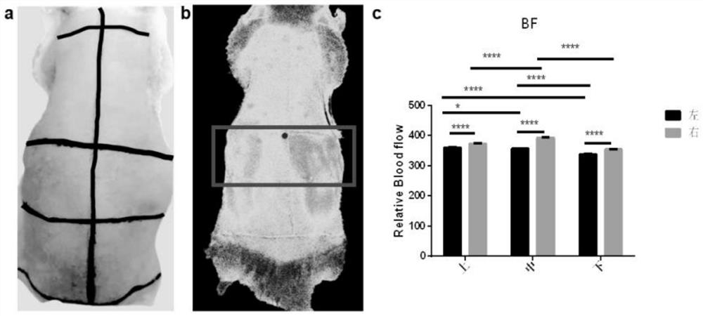
[0057] The laser speckle blood flow imaging unit uses a laser speckle blood flow imager (model: SIM BFI HR Pro) for macroscopic observation of blood vessels and blood perfusion.

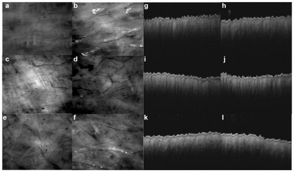
[0058] The two-photon imaging unit includes a two-photon confocal scanning microscope (Olympus FV1000) and a Mai TaiDeepSee laser (120fs, 80MHz) for microscopic observation of vessel shape and shape;

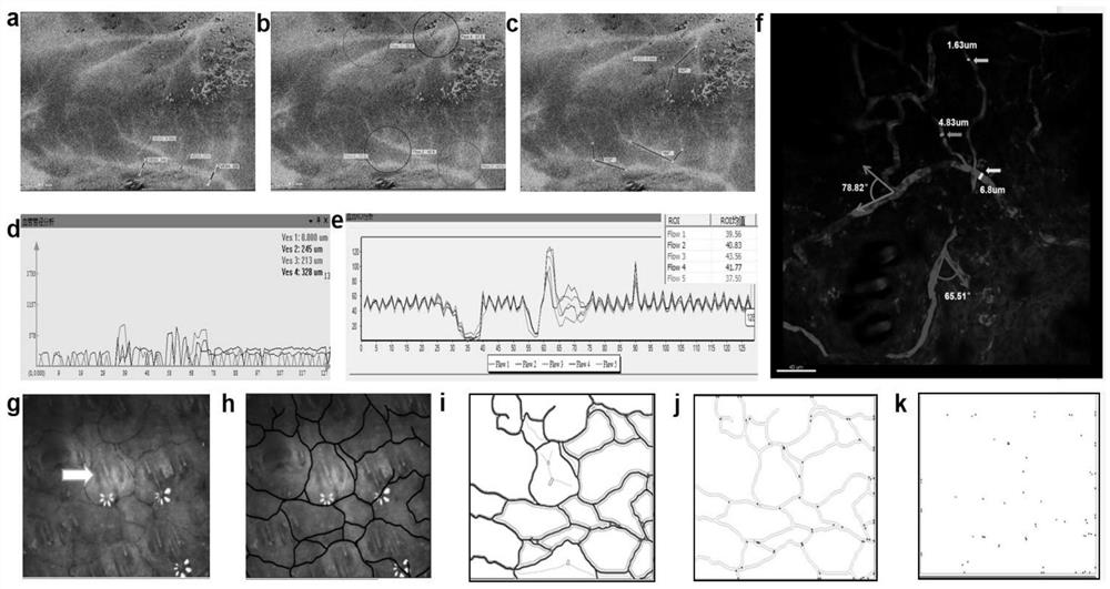
[0059] The capillary detection unit selects the capillary detector (model: CAM1CVF) for the observation of the capillary network;

[0060]The immunofluorescence staining unit includes section preparation materials, immunofluorescence reaction materials and fluorescence observation equipment, which are used for counting transverse and longitudinal blood vessels at different d...

Embodiment 2

[0063] The system of Example 1 was used to evaluate skin vessels and blood flow.

[0064] 1. Observation location selection

[0065] (1) Specklemeter observation

[0066] Three male Wistar rats with a body weight of 190±20 g were selected.

[0067] Rats were anesthetized with isoflurane, and most of the back hair was shaved with a shaver, then the remaining hair was cleaned with depilatory cream, and finally wiped with warm saline. Keep the rats in a state of continuous anesthesia, put them under the macroscopic microscope of the laser speckle blood flow imager, observe the skin on the back, and then connect the computer images to perform real-time positioning calibration, and collect real-time images and blood flow images at the same time. When the images are relatively clear, the image acquisition is carried out, and the acquisition time is 100s.

[0068] Divide its back into six areas, namely the upper, middle, and lower areas, and each area is evenly divided into left a...


PUM

PropertyMeasurementUnit
Diameteraaaaaaaaaa
Diameteraaaaaaaaaa
Diameteraaaaaaaaaa

Abstract

The invention discloses a skin blood vessel and blood flow quantitative evaluation system and method based on optical imaging, and belongs to the technical field of biological information processing. The skin blood vessel and blood flow quantitative evaluation system based on optical imaging comprises a laser speckle blood flow imaging unit, a two-photon imaging unit, a blood capillary detection unit and an immunofluorescence staining unit. The laser speckle blood flow imaging unit, the blood capillary detection unit and the immunofluorescence staining unit are used for macroscopic observation, the two-photon imaging unit is used for microscopic morphology observation, observation results of all the units are synthesized, establishment of a corium blood vessel and blood flow quantitative evaluation system is achieved, and a powerful guarantee is provided for research of a blood vessel blood flow microcirculation system.

Description

technical field [0001] The invention belongs to the technical field of biological information processing, and in particular relates to a system and method for evaluating skin blood vessels and blood flow quantification based on optical imaging. Background technique [0002] The skin microcirculation vascular network has the characteristics of three-dimensional construction. The hairpin-shaped capillary loops on the outermost layer are located in the dermal papilla, and each papilla has one or more loops. It has a hedgehog skin-like appearance as a whole; there is a layer of polygonal capillary network characterized by a polygonal grid under the papilla at the junction of the papillary layer of the dermis and the reticular layer. The rich vascular network is the surrounding nerves, hair follicles, and glands It is very important to provide adequate nutrition. [0003] At present, the observation of the morphology of dermal blood vessels and the amount of blood perfusion is s...

Claims


Application Information

Patent Timeline
IPC IPC(8): A61B5/026A61B5/0275A61B5/02A61B5/00
CPCA61B5/0261A61B5/02007A61B5/0275A61B5/72A61B2503/40A61B2503/42
Inventor 孙娅楠李俐霜王毅
Owner EXPERIMENTAL RES CENT CHINA ACAD OF CHINESE MEDICAL SCI
Why Eureka
Try Eureka