Intraoperative conjunctival sac drainage apparatus for ophthalmologic operation

An ophthalmic surgery and conjunctival sac technology, applied in the directions of surgery, suction equipment, medical science, etc., can solve the problems of wrong surgical operation, affecting the normal operation of the operator, and heavy workload, and achieve the effect that is not easy to hinder

Pending Publication Date: 2022-04-15
THE EYE HOSPITAL OF WENZHOU MEDICAL UNIV
View PDF0 Cites 0 Cited by
  • Summary
  • Abstract
  • Description
  • Claims
  • Application Information

AI Technical Summary

Problems solved by technology

Because the cornea is exposed for a long time during the operation, it is necessary to moisten the cornea frequently with balanced salt solution to ensure the cornea is transparent and the surgical field is clear enough. The conjunctival sac of such patients is deep, and the balanced salt solution can easily accumulate in the conjunctival sac during the operation, and even submerge the cornea, resulting in unclear operative field and seriously affecting the operation of the surgeon.
[0003] At present, in clinical practice, assistants usually use cotton swabs or gauze to drain or suck out the effusion in time. In order to ensure a continuous clear operation field, assistants are required to clean up the effusion frequently during the operation. The effusion operation will hinder the operator and affect the normal operation of the operator. If the cooperation is not good, it may lead to surgical operation errors and cause serious consequences

Method used


Image

  • Intraoperative conjunctival sac drainage apparatus for ophthalmologic operation
  • Intraoperative conjunctival sac drainage apparatus for ophthalmologic operation
  • Intraoperative conjunctival sac drainage apparatus for ophthalmologic operation

Examples


Embodiment 1

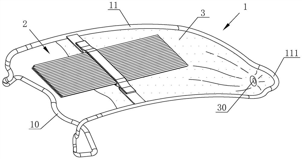
[0029] Such as Figure 1 to Figure 4 The shown conjunctival sac drainage device for ophthalmic surgery includes a suction mechanism 2 for draining conjunctival sac fluid and a fixing bracket 1 for fixing the suction mechanism 2 at the corner of the eye. The fixing bracket 1 includes a The inner eyelid fixing frame 10 and the extension frame 11 are made of medical grade stainless steel. The lower eyelid embedding part 101, the eye corner relief part 102 and two transition parts 103 for connecting the extension frame 11 are composed. The eyelid embedding part 101 and the eye corner relief part 102 are located between the upper eyelid embedding part 100 and the lower eyelid embedding part 101 and are curved in an arc. The upper eyelid embedding part 100 and the lower eyelid embedding part 101 can respectively extend into the inner side of the upper and lower eyelids close to the outer corner of the eye, so as to maintain the fixation of the fixing bracket 1 and open the corner o...

Embodiment 2

[0036] Such as Figure 5-7 As shown, the technical content of this embodiment is basically the same as that of Embodiment 1, and the technical points of difference between this embodiment and Embodiment 1 are:

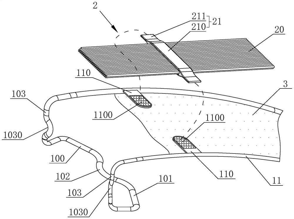
[0037] The sliver fixing plate 210 is provided with a cover plate 2101 for opening and closing the fixing groove 2100, one end of the cover plate 2101 can be turned up and down through a hinged structure, and the other end of the cover plate 2101 can be locked and fixed on the sliver fixing plate at the end of the fixing groove 2100 210 , the inner wall of the fixing groove 2100 is distributed with a plurality of insertion rods 2102 for preventing the liquid-absorbing sliver 20 from slipping off. The liquid-absorbing sliver 20 can be replaced, so that the sliver fixing plate 210 can be recycled with the fixed bracket 1. When the liquid-absorbing sliver 20 is installed, the insertion rod 2102 can be inserted into the liquid-absorbing sliver 20 to adjust the liquid-absor...

Embodiment 3

[0039] Such as Figure 8 with Figure 9 As shown, the technical content of this embodiment is basically the same as that of Embodiment 1, and the technical points of difference between this embodiment and Embodiment 1 are:

[0040] The eyelid fixing frame 10 includes an upper eyelid stretcher 104 and a lower eyelid stretcher 105 which are independent and symmetrically arranged, and the extension frame 11 is a "U"-shaped frame, and both ends of the extension frame 11 are bent inwards at the same time. The lower bend finally connects the upper eyelid stretcher 104 and the lower eyelid stretcher 105 respectively, the docking plate 110 includes a first half plate 112 and a second half plate 113, and the end of the upper eyelid stretcher 104 is bent upward and fixedly connected The first half plate 112 and the end of the lower eyelid stretching portion 105 are bent upwards and fixedly connected to the second half plate 113 . Since the upper and lower eyelid stretchers 105 are rel...


PUM


Abstract

The invention discloses an intraoperative conjunctival sac drainage apparatus for ophthalmologic operation, which can be placed at the outer canthus of a patient during the operation, adopts a flexible suction structure to directly contact with effusion in the conjunctival sac, and can continuously suck the effusion outwards according to capillary action to keep the surgical field clear. According to the technical scheme, the device is characterized by comprising a suction mechanism used for leading out conjunctival sac effusion and a fixing support used for fixing the suction mechanism to the canthus, the suction mechanism comprises a flexible suction structure capable of making direct contact with the ocular surface, and the flexible suction structure sucks the conjunctival sac effusion through the capillary action; the flexible attraction structure is in butt joint and fixed with the fixed bracket through the fixed butt joint piece; the fixing support comprises an eyelid fixing frame capable of extending into the inner side of an eyelid and an extension frame, the eyelid fixing frame and the extension frame are integrally formed, the extension frame is provided with a drainage film, one end of the flexible suction structure makes contact with conjunctival sac effusion, the other end of the flexible suction structure is laid on the drainage film, and the drainage film is used for collecting or discharging the effusion led out by the flexible suction structure.

Description

technical field [0001] The invention relates to the field of equipment related to ophthalmic surgery, in particular to a conjunctival sac drainage device during ophthalmic surgery. Background technique [0002] Ophthalmic surgery is a delicate operation, and it is particularly important to ensure a clear vision during the operation. Because the cornea is exposed for a long time during the operation, it is necessary to moisten the cornea frequently with balanced salt solution to ensure the cornea is transparent and the surgical field is clear enough. The conjunctival sac of such patients is deep, and the balanced salt solution can easily accumulate in the conjunctival sac during the operation, and even submerge the cornea, resulting in unclear operative field and seriously affecting the operation of the surgeon. [0003] At present, in clinical practice, assistants usually use cotton swabs or gauze to drain or suck out the effusion in time. In order to ensure a continuous cl...

Claims


Application Information

Patent Timeline
IPC IPC(8): A61M1/00A61B17/02
Inventor 许淑霞梁远波张绍丹李国星
Owner THE EYE HOSPITAL OF WENZHOU MEDICAL UNIV
Why Eureka
Try Eureka