
Image display method, computer equipment and storage medium

A technology of image display and computer program, which is applied in the field of medical image processing, can solve the problems of low efficiency of analysis methods, and achieve the effect of improving efficiency and accuracy

Pending Publication Date: 2022-07-01
SHANGHAI UNITED IMAGING HEALTHCARE
View PDF0 Cites 0 Cited by
  • Summary
  • Abstract
  • Description
  • Claims
  • Application Information

AI Technical Summary

Problems solved by technology

[0004] However, for plaque analysis, the common clinical solution is to analyze and evaluate the overall vessel morphology on the 3D sequence, or to perform plaque detail analysis on the 2D sequence, which is extremely inefficient.

Method used


Image

  • Image display method, computer equipment and storage medium
  • Image display method, computer equipment and storage medium
  • Image display method, computer equipment and storage medium

Examples


Embodiment Construction

[0056] In order to make the purpose, technical solutions and advantages of the present application more clearly understood, the present application will be described in further detail below with reference to the accompanying drawings and embodiments. It should be understood that the specific embodiments described herein are only used to explain the present application, but not to limit the present application.

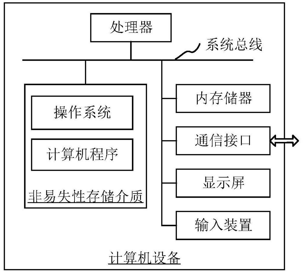
[0057] The image display method provided in this application can be applied to such as figure 1 In the computer equipment shown, the computer equipment can be a server, and the computer equipment can also be a terminal, and its internal structure diagram can be as follows: figure 1shown. The computer equipment includes a processor, memory, a network interface, a display screen, and an input device connected by a system bus. Among them, the processor of the computer device is used to provide computing and control capabilities. The memory of the computer device includ...


PUM


Abstract

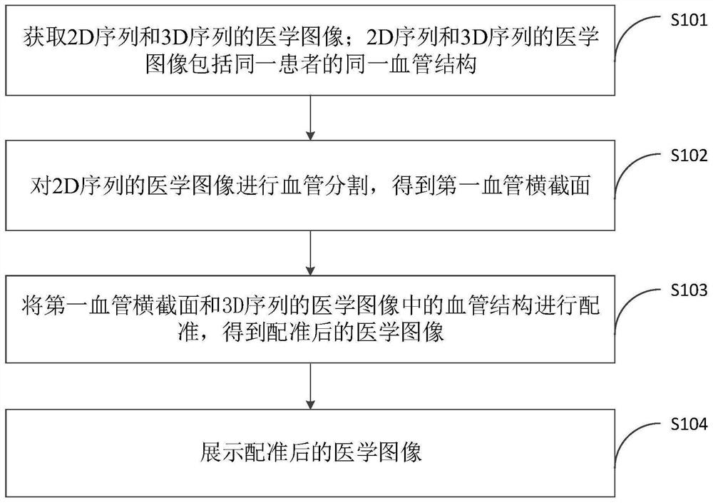
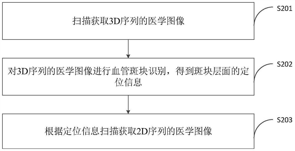
The invention relates to an image display method, computer equipment and a storage medium. The method comprises the following steps: acquiring medical images of a 2D sequence and a 3D sequence of the same vascular structure of the same patient, performing vascular segmentation on the medical images of the 2D sequence to obtain a first vascular cross section, and registering the first vascular cross section and the vascular structure in the medical images of the 3D sequence to obtain a registered medical image. According to the method, the first blood vessel cross section of the 2D sequence and the blood vessel structure of the 3D sequence are displayed on the same display interface at the same time, the whole blood vessel structure is displayed from the 3D dimension, the blood vessel cross section is also displayed from the 2D level, the overall form and local information of the blood vessel structure are included, and the display effect is good. And sufficient and reliable reference data is provided for a doctor to carry out plaque analysis based on the images of the 3D and 2D sequences displayed on the display interface, so that the accuracy of plaque analysis is improved.

Description

technical field [0001] The present application relates to the technical field of medical image processing, and in particular, to an image display method, computer equipment and storage medium. Background technique [0002] High-resolution magnetic resonance imaging (HR-MRI) technology is one of the commonly used techniques for clinical diagnosis of carotid plaques. It has high spatial resolution and tissue contrast. In recent years, more and more studies have applied HR-MRI to diagnose and analyze carotid artery plaque features, such as intraplaque hemorrhage, fibrous cap, and lipid core. High-resolution "black blood" technology and "bright blood" 3D-TOF (3Dtime-of-flight) technology can not only increase the contrast between the wall and lumen, but also obtain information on the morphology and composition of carotid plaques, including Measure the total volume of the plaque, identify the components of the plaque (such as intraplaque hemorrhage, fibrous cap, lipid core, etc...

Claims


Application Information

Patent Timeline
Patent Type & Authority Applications(China)
IPC IPC(8): G06T7/10G06T7/30G06T7/70
CPCG06T7/10G06T7/30G06T7/70G06T2207/10012G06T2207/30101
Inventor 苏赛赛任晓敏滕艳群
Owner SHANGHAI UNITED IMAGING HEALTHCARE
Learn More