Processing system and device for fluorescein fundus angiography image

A technology of angiography and fluorescein, applied in the field of computer vision, can solve the problems of poor applicability, unrealized lesion quantification, inability to treat retinal lesions, etc., and achieve good applicability

Active Publication Date: 2022-07-22
ZHONGSHAN OPHTHALMIC CENT SUN YAT SEN UNIV
View PDF6 Cites 1 Cited by
  • Summary
  • Abstract
  • Description
  • Claims
  • Application Information

AI Technical Summary

Problems solved by technology

However, in the prior art, only the segmentation and positioning of the non-perfusion area in diabetic retinopathy (DR) has not been realized, and the quantification of the lesion has not been realized, and the problem of poor applicability is that it cannot be processed for other retinopathy

Method used


Image

  • Processing system and device for fluorescein fundus angiography image
  • Processing system and device for fluorescein fundus angiography image
  • Processing system and device for fluorescein fundus angiography image

Examples


Embodiment 1

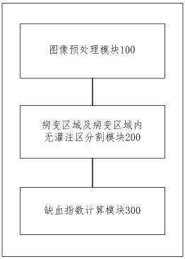
[0051] See figure 1 , an embodiment of the present application provides a fluorescein fundus angiography image processing system, the system includes:

[0052] The image preprocessing module 100 is configured to obtain a fluorescein fundus angiography image to be processed, and preprocess the fluorescein fundus angiography image, where the fluorescein fundus angiography image is any one of preset retinal lesions. Fluorescein angiography image of the lesion.

[0053] Among them, the fluorescein fundus angiography image to be processed can be a 55-degree fluorescein fundus angiography image (FFA image); the preset multiple retinopathy can include diabetic retinopathy (DR), branch retinal vein occlusion (BRVO) At least two of , central retinal vein occlusion (CRVO), and retinal vasculitis, therefore, the fluorescein fundus angiography image to be processed may be an FFA image of a patient with any of the above-mentioned retinopathy.

[0054] Specifically, the image preprocessin...

Embodiment 2

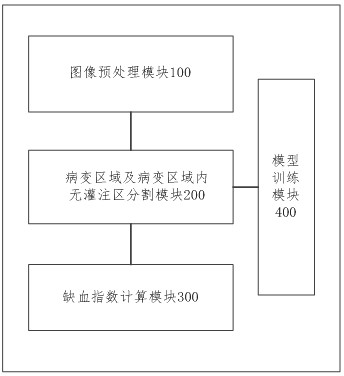
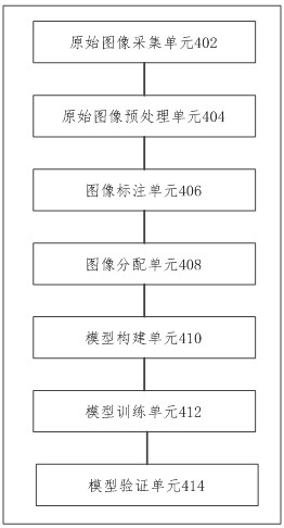
[0066] Based on the first embodiment above, in this embodiment, please refer to figure 2 and image 3 , the system also includes a model training module 400, the model training module 400 includes:

[0067] The original image acquisition unit 402 is used for acquiring original images to create an original image set.

[0068] The original image may be an FFA image of DR or BRVO, and the original image set includes FFA images of DR and BRVO.

[0069] Specifically, the original image acquisition unit 402 acquires a 55-degree FFA image diagnosed as DR or BRVO as an original image, and creates an original image set based on a large number of acquired original images.

[0070] The original image preprocessing unit 404 is configured to preprocess each original image in the original image set, and delete the original images that do not meet the preset standard from the original image set to obtain a preprocessed image set.

[0071] The preset standard may be a preset model input i...

Embodiment 3

[0091] Based on the second embodiment above, in this embodiment, please refer to Figure 4 , the system further includes an image display module 500 .

[0092] The image display module 500 can be used to display the fluorescein fundus angiography image, and use different designated colors to display the lesion area and the non-perfusion area in the fluorescein fundus angiography image, so as to realize the intuitive display of the non-perfusion area, which is helpful for Assisting doctors in determining the specific location of laser treatment and implementing precision medicine can also simplify laser surgery and promote the ability of grassroots hospitals to carry out laser treatment; The area value of the perfusion area and the clinical ischemia index corresponding to the fluorescein fundus angiography image can automatically display the area value of the diseased area of ​​the patient's retina, the area value of the non-perfusion area and the clinical ischemia index, which...


PUM


Abstract

The invention belongs to the technical field of computer vision, and discloses a fluorescein fundus angiography image processing system and device, and the system comprises an image preprocessing module which is used for obtaining a to-be-processed fluorescein fundus angiography image, and carrying out the preprocessing of the fluorescein fundus angiography image; the lesion area and lesion area non-perfusion area segmentation module is used for processing the preprocessed fluorescein fundus angiography image through a pre-trained semantic segmentation model, and determining a lesion area in the image and a non-perfusion area in the lesion area; and the ischemic index calculation module is used for acquiring the area value of the lesion region and the area value of the non-perfusion region, and calculating to obtain the clinical ischemic index corresponding to the fluorescein fundus angiography image. According to the method, the lesion area in the fluorescein fundus angiography image and the non-perfusion area in the lesion area can be segmented, lesion quantification is realized, and the method is suitable for various retinopathy effects.

Description

technical field [0001] The present application relates to the technical field of computer vision, and in particular, to a system and device for processing fluorescein fundus angiography images. Background technique [0002] There are many classifications of retinopathy, such as diabetic retinopathy (DR), branch retinal vein occlusion (BRVO), central retinal vein occlusion (CRVO) and retinal vasculitis; fluorescence Fundus Fluorescein Angiography (FFA) can reflect retinal blood vessels and perfusion. However, the existing technology only targets the segmentation and localization of the non-perfusion area in diabetic retinopathy (DR), which not only fails to quantify the lesions, but also cannot be processed for other retinopathy, and has poor applicability. SUMMARY OF THE INVENTION [0003] The present application provides a fluorescein fundus angiography image processing system and device, which can segment the lesion area in the fluorescein fundus angiography image and t...

Claims


Application Information

Patent Timeline
Patent Type & Authority Applications(China)
IPC IPC(8): G06T7/00G06T7/62G06V40/18G06V10/26G06V10/82G06N3/04G16H50/20
CPCG06T7/0012G06T7/62G16H50/20G06T2207/10064G06T2207/30041G06T2207/30101G06N3/045
Inventor 林桢哲林铎儒于珊珊林浩添梁小玲赵欣予
Owner ZHONGSHAN OPHTHALMIC CENT SUN YAT SEN UNIV
Why Eureka
Try Eureka