Integrated electromyographic clinician programmer for use with an implantable neurostimulator

a neurostimulator and electromyographic technology, applied in the field of neurostimulator treatment systems, can solve the problems of difficult to accurately predict or identify specific organs, difficult to determine the viability of treatment, and significant disadvantages of current stimulation electrode placement/implantation techniques and known treatment setting techniques, so as to improve the consistency of outcomes, reduce the duration and complexity of the procedure, and improve the positioning or programming of the implantable lead.

Active Publication Date: 2018-10-09
AXONICS
View PDF598 Cites 13 Cited by
  • Summary
  • Abstract
  • Description
  • Claims
  • Application Information

AI Technical Summary

Benefits of technology

[0009]The integrated EMG clinician programmer of the present invention provides an objective and quantitative means by which to standardize placement and programming of implantable leads and neurostimulation electrodes, reducing the subjective assessment of patient sensory responses as well as surgical, programming, and re-programming time. Further, as the efficacy of treatment often relies on precise placement of the neurostimulation electrodes at target tissue locations and the consistent, repeatable delivery of neurostimulation therapy, using an objective EMG measurement can substantially improve the utility and success of treatment. Use of the integrated EMG clinician programmer to verify activation of motor responses can further improve the lead placement performance of less experienced operators and allow such physicians to perform lead placement with confidence and greater accuracy. Still further, automation of several steps or procedures associated with lead placement and programming with the integrated clinician programmer can further reduce the duration and complexity of the procedure and improve consistency of patient outcomes. For example, automation of electrode threshold determinations based on EMG responses can provide rapid feedback during lead placement and to identify optimal programming parameters.
[0010]An integrated electromyography (EMG) and signal generation clinician programmer may be coupled with an implantable temporary or permanent lead in a patient and at least one EMG sensing electrode minimally invasively positioned on a skin surface or within the patient. Generally, the integrated clinician programmer may comprise a portable housing, a signal / stimulation generator, an EMG signal processor / recorder, and a graphical user interface. The housing has an external surface and encloses circuitry at least partially disposed within the housing. The signal / stimulation generator may be disposed within the housing and configured to deliver test stimulation to a nerve tissue of the patient via a percutaneous needle or the implantable lead. The EMG signal processor may be disposed within the housing and configured to record a stimulation-induced EMG motor response for each test stimulation via the at least one pair of EMG sensing electrodes and a ground electrode. The graphical user interface at least partially comprises the external surface of the housing and has a touch screen display for direct user interaction or for use with a keyboard, mouse, or the like. As described in greater detail below, the integrated clinician programmer allows for controlled positioning or programming of the implantable lead based at least on the EMG record and provides the clinician with a convenient all-in-one setup via the EMG integrated clinician programmer.
[0011]The graphical user interface of the integrated clinician programmer may include an EMG display comprising a visual image of the EMG record, wherein the visual image includes a waveform comprising a compound muscle action potential (CMAP) and / or a visual bar to indicate a maximum CMAP response (e.g., maximum peak, peak to peak). The EMG display may further include a motor response graphical element which is configured for user input of the EMG motor response (e.g., yes, no) associated with each test stimulation. The graphical user interface may also include a sensory response graphical element which is configured for user input of a sensory response (e.g., none, good, bad) from the patient associated with each test stimulation. As discussed in greater detail below, user characterization of the presence or absence of motor and / or sensory responses may be of additional benefit in fine tuning lead placement.
[0012]The graphical user interface includes a stimulation amplitude adjustment graphical element which is configured for user adjustment of a stimulation amplitude of the test stimulation from the signal generator in increments in a range from about 0.05 mA to about 0.25 mA, wherein the test stimulation amplitude is generally less than 10 mA. The use of proportional increases in stimulation amplitude during test stimulation and / or programming effectively reduces the time required for such activities. The graphical user interface may further include at least one parameter graphical element which is configured for user adjustment of a pulse width of the test simulation, a pulse frequency of the test stimulation, a cycling or continuous mode of the test stimulation, a bipolar or monopolar mode of the test stimulation, or an electrode configuration of the implantable lead.
[0018]The present invention further provides for automated methods for improved positioning or programming of an implantable lead in a patient with an integrated electromyography (EMG) and signal generation clinician programmer coupled to the implantable lead. At least one EMG sensing electrode is minimally invasively positioned on a skin surface or within the patient and coupled to the integrated clinician programmer. The method comprises generating a test stimulation from the integrated clinician programmer and delivering the test stimulation to a nerve tissue of the patient with the implantable lead. A stimulation-induced EMG motor response is detected with the integrated clinician programmer for each test stimulation via the least one EMG sensing electrode. A stimulation amplitude of the test stimulation is automatically adjusted and the delivering and detecting steps are repeated until a desired stimulation-induced EMG motor response is detected. As discussed above, automation of certain aspects within the clinician programmer can further reduce the duration and complexity of the procedure and improve consistency of outcomes. In this instance, the clinician programmer is configured with an automated threshold determination based on EMG responses to provide rapid feedback during lead placement and to identify optimal programming parameters.
[0019]The desired EMG motor response may comprise a value associated with a minimum or maximum compound muscle action potential (CMAP). Automatically adjusting may comprise increasing the stimulation amplitude in increments of 0.05 mA for a test stimulation less than or equal to 1 mA, 0.1 mA for a test stimulation more than or equal to 1 mA and less than or equal to 2 mA, 0.2 mA for a test stimulation more than or equal to 2 mA and less than or equal to 3 mA, or 0.25 mA for a test stimulation more than or equal to 3 mA. The use of proportional increases in combination with the automated feature in stimulation amplitude adjusting during test stimulation and / or programming effectively reduces the time required for such activities. It will be appreciated that this automated feature may be easily terminated at any time and for any reason, patient safety or otherwise, by the user.

Problems solved by technology

A number of factors may lead to the very different outcomes that patients experience, and viability of treatment can be difficult to determine before implantation.
Current stimulation electrode placement / implantation techniques and known treatment setting techniques suffer from significant disadvantages.
The nerve tissue structures of different patients can be quite different, with the locations and branching of nerves that perform specific functions and / or enervate specific organs being challenging to accurately predict or identify.
The electrical properties of the tissue structures surrounding a target nerve structure may also be quite different among different patients, and the neural response to stimulation may be markedly dissimilar, with an electrical stimulation pulse pattern, pulse width, frequency, and / or amplitude that is effective to affect a body function of one patient and potentially imposing significant discomfort or pain, or having limited effect, on another patient.
Even in patients where implantation of a neurostimulation system provides effective treatment, frequent adjustments and changes to the stimulation protocol are often required before a suitable treatment program can be determined, often involving repeated office visits and significant discomfort for the patient before efficacy is achieved.
While a number of complex and sophisticated lead structures and stimulation setting protocols have been implemented to seek to overcome these challenges, the variability in lead placement results, the clinician time to establish suitable stimulation signals, and the discomfort (and in cases the significant pain) that is imposed on the patient remain less than ideal.
In addition, the lifetime and battery life of such devices is relatively short, such that implanted systems are routinely replaced every few years, which requires additional surgeries, patient discomfort, and significant costs to healthcare systems.
Furthermore, since the morphology of the nerve structures vary considerably between patients, placement and alignment of neurostimulation leads relative the targeted nerve structures can be difficult to control, which can lead to inconsistent placement, unpredictable results and widely varying patient outcomes.
One drawback with this approach is that repeated office visits may be required to determine the appropriate electrodes to use and / or to arrive at a neurostimulation program that delivers effective treatment.
Often, the number of usable neurostimulation programs may be limited by imprecise lead placement.
The tremendous benefits of these neural stimulation therapies have not yet been fully realized.

Method used


Image

  • Integrated electromyographic clinician programmer for use with an implantable neurostimulator
  • Integrated electromyographic clinician programmer for use with an implantable neurostimulator
  • Integrated electromyographic clinician programmer for use with an implantable neurostimulator

Examples


Embodiment Construction

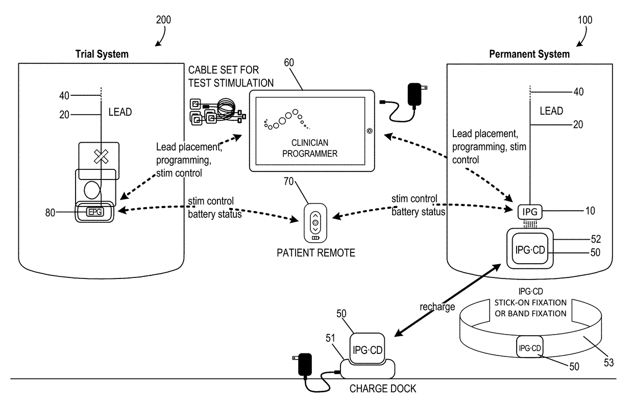
[0043]The present invention relates to neurostimulation treatment systems and associated devices, as well as methods of treatment, implantation / placement and configuration of such treatment systems. In particular embodiments, the invention relates to sacral nerve stimulation treatment systems configured to treat bladder dysfunctions, including overactive bladder (“OAB”), as well as fecal dysfunctions and relieve symptoms associated therewith. For ease of description, the present invention may be described in its use for OAB, it will be appreciated however that the present invention may also be utilized for any variety of neuromodulation uses, such as bowel disorders (e.g., fecal incontinence, fecal frequency, fecal urgency, and / or fecal retention), the treatment of pain or other indications, such as movement or affective disorders, as will be appreciated by one of skill in the art.

I. Neurostimulation Indications

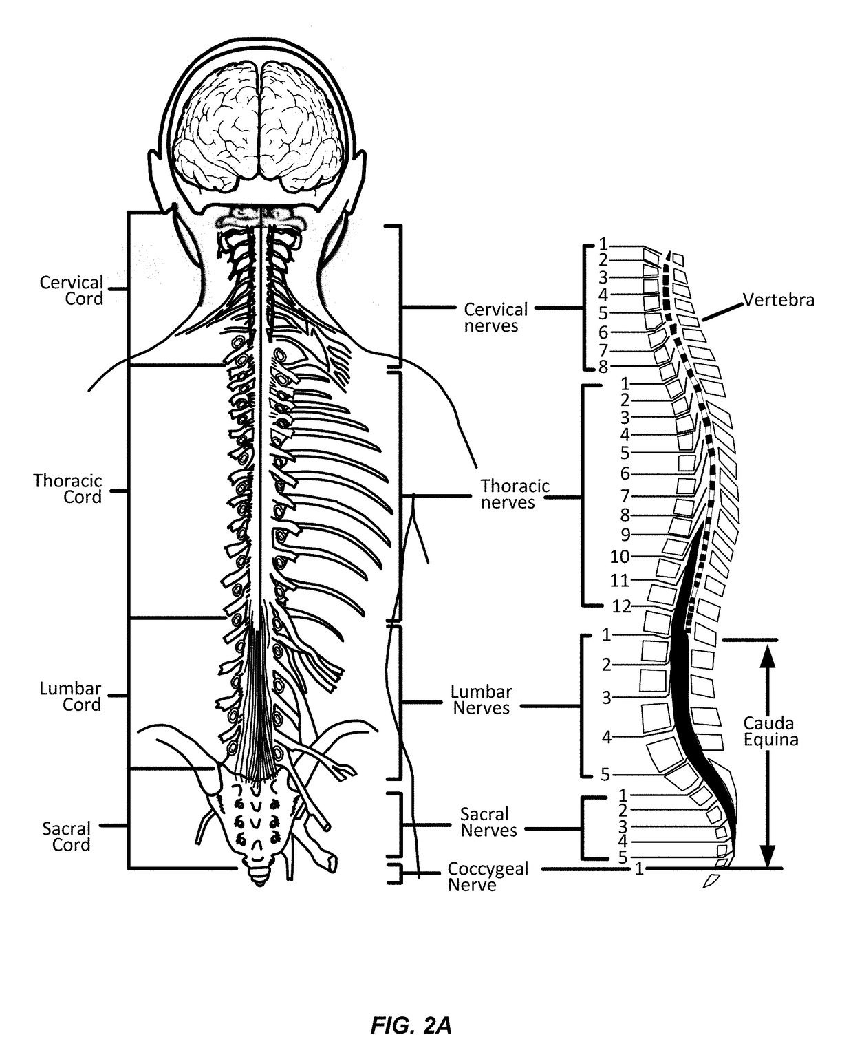
[0044]Neurostimulation (or neuromodulation as may be used interchangeabl...


PUM


Abstract

An integrated electromyography (EMG) and signal / stimulation generation clinician programmer may be coupled with an implantable temporary or permanent lead in a patient and at least one EMG sensing electrode minimally invasively positioned on a skin surface or within the patient. Generally, the integrated clinician programmer may comprise a portable housing, a signal / stimulation generator, and EMG signal processor, and a graphical user interface. The signal / stimulation generator may be disposed within the housing and configured to deliver test stimulation to a nerve tissue of the patient via the implantable lead. The EMG signal processor may be disposed within the housing and configured to record a stimulation-induced EMG motor response for each test stimulation via the at least one EMG sensing electrode. The graphical user interface at least partially comprises the external surface of the housing and has a touch screen display for direct user interaction or a keyboard, mouse, or the like.

Description

CROSS-REFERENCES TO RELATED APPLICATIONS[0001]The present application is a Non-Provisional of and claims the benefit of priority of U.S. Provisional Application Nos. 62 / 038,131 filed Aug. 15, 2014; 62 / 041,611 filed Aug. 25, 2014; and 62 / 101,899 filed Jan. 9, 2015; each of which is incorporated herein by reference in its entirety for all purposes.[0002]The present application is related to concurrently filed U.S. Non-Provisional patent application Ser. No. 14 / 827,074, entitled “Devices and Methods for Anchoring of Neurostimulation Leads”; U.S. Non-Provisional patent application Ser. No. 14 / 827,081, entitled “External Pulse Generator Device and Associated Methods for Trial Nerve Stimulation”; U.S. Non-Provisional patent application Ser. No. 14 / 827,108, entitled “Electromyographic Lead Positioning and Stimulation Titration in a Nerve Stimulation System for Treatment of Overactive Bladder”; and U.S. Non-Provisional patent application Ser. No. 14 / 827,067, entitled “Systems and Methods fo...

Claims


Application Information

Patent Timeline
Patent Type & Authority Patents(United States)
IPC IPC(8): A61N1/372A61N1/36A61N1/05A61B5/296
CPCA61N1/37241A61N1/36007A61N1/36017A61N1/37247A61N1/36185A61N1/36107A61N1/36139A61N1/36157A61N1/0551A61N1/3603
Inventor JIANG, GUANGQIANGWOOCK, JOHNSCHROEDER, DENNISSCHMID, ERIC
Owner AXONICS
Why Eureka
Try Eureka