Automated cell and tissue bioprinter

a bioprinter and tissue technology, applied in the field of tissue engineering, can solve problems such as the limitation of cell placement and alignment, and achieve the effects of reducing the width of the pattern, reducing the size, and increasing the speed

Active Publication Date: 2018-11-06
THE UNIVERSITY OF AKRON
View PDF7 Cites 3 Cited by
  • Summary
  • Abstract
  • Description
  • Claims
  • Application Information

AI Technical Summary

Benefits of technology

[0006]One or more embodiments of the present invention are directed to an automated mechanism and related methods for making three-dimensional tissue constructs with spatial organization of cells that provides: (i) organized cell placement and spatial assembly of multiple cell types in a reproducible manner (ii) direct and non-contact assembly of multiple cell types and / or cell layers without exerting damaging forces on the cells or tissues, (iii) retention of cell viability and functionality during assembly steps and long periods of culture, (iv) minimal use of harmful and toxic chemicals such as profuse amounts of mineral oil and buffer reagent, and (v) efficient layer-to-layer assembly of cell layers within the three-dimensional construct while (vi) avoiding the use of complicated surface treatments that prevents scale-up, and (vii) avoiding mutation-inducing radiations such as UV used to photocrosslink hydrogels.

Problems solved by technology

A major challenge in the field of tissue engineering is creating two- and three-dimensional multi-cellular constructs with defined spatial organization of cells, to mimic the architecture of living tissues, where homotypic and heterotypic cellular interactions are key to tissue formation and function.
Although both classes show some promise, bottom-up approaches can potentially overcome limitations in cell placement and alignment that are difficult to achieve in most top-down approaches.
Unfortunately, these systems showed only the capability to print bioreagents and cells in arbitrary shapes, because printing was performed with manual liquid handling tools, it could only control droplet-shaped patterns.

Method used


Image

  • Automated cell and tissue bioprinter
  • Automated cell and tissue bioprinter
  • Automated cell and tissue bioprinter

Examples


example 1

Preparation of Aqueous PEG and DEX Stock Solutions

[0094]For all printing experiments, an ATPS with 5.0% (w / w) polyethylene glycol (PEG, Mw: 35 k) and 6.4% (w / w) dextran (DEX, Mw: 500 k), was used as phase-forming polymers. PEG and DEX were obtained from Sigma and Pharmacosmos, respectively. Each phase was individually prepared with these concentrations in growth medium. The DEX phase was prepared twice the final concentration to account for mixing with the PEG and cell suspension at a 1:1 ratio (FIG. 8A). Centrifuge tubes containing the solutions were allowed to rest in a vertical position at room temperature for ˜2 hours for the polymer to fully dissolve. The stock solutions were then stored at 4° C.

example 2

Preparation of Printing Surface and Printing Phase

[0095]For each experiment, the printing surface was maintained in 5% (w / w) PEG (FIG. 8B). Five different types of surfaces were prepared or provided to assess the stability of printed patterns of the DEX phase, including decellularized matrices, cell monolayer replica, live cell monolayers of different confluence, molecularly smooth films of poly(D,L-lactic acid) (PDLA), and PDLA with fabricated striped microgrooves of 10 and 20 μm pitch.

[0096]Preparation of each surface is briefly discussed: (i) Decellularized matrices of MDA-MB-231 breast cancer cells and C2C12 mouse myoblast cells were prepared by treating a confluent monolayer with a 0.25% (v / v) Triton X-100 (Sigma) solution in PBS for 15 minutes. The monolayer was then washed three times with PBS to ensure the complete removal of all cell constituents except for the protein matrix laid down by cells on the surface. When prepared at least a day before printing, samples were maint...

example 3

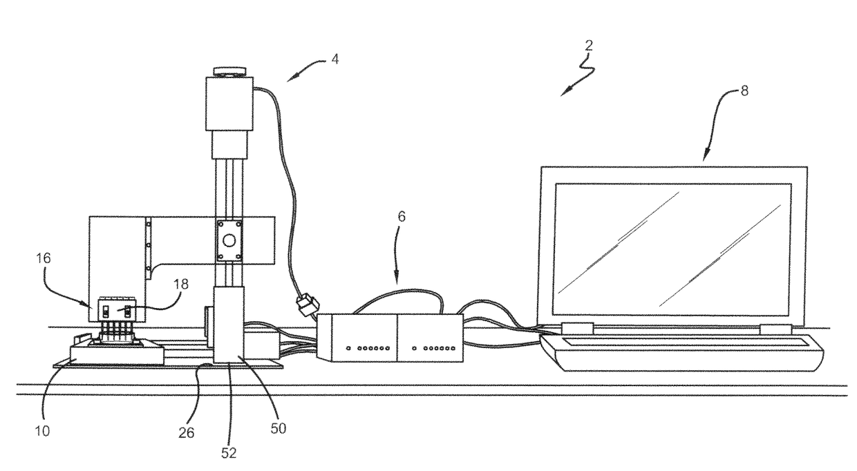
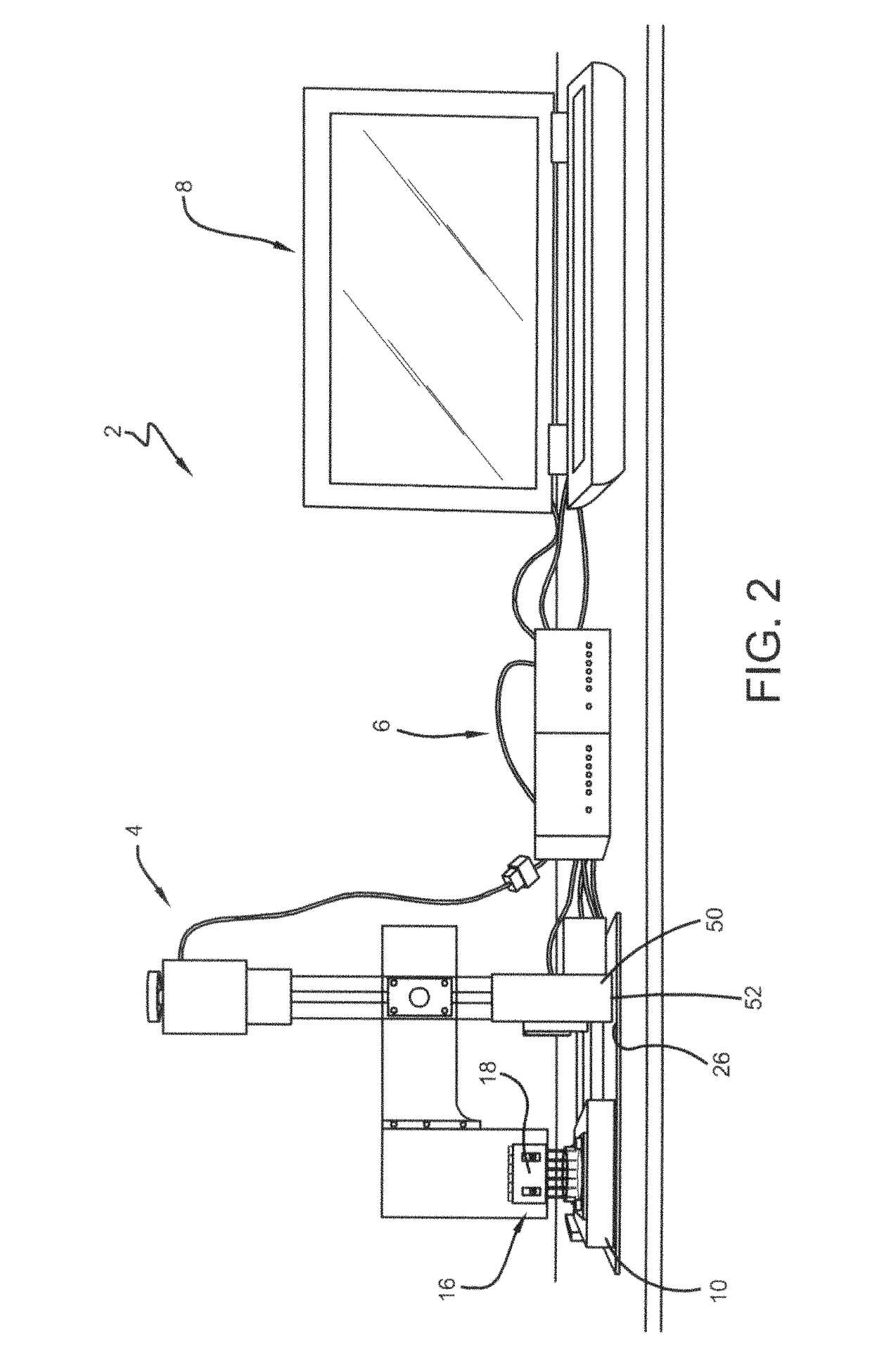
Calibrations of the Three-Axis Motorized System

[0101]The three-axis motorized system was calibrated at the beginning of each experiment. The printer head containing the cartridge and printing tips was programmed to descend into an empty dish until the tip touched the dish surface. The printer head was then programmed to ascend ˜500 μm (78 steps) to prevent tips from scraping the printed surface during printing. Another script of commands was developed to print individual patterns for characterization studies. The printing tips were programmed to descend into the dish (FIG. 8C), perform a linear forward motion, ascend out of the dish, and then move an offset distance to prepare for the next print. The offset distances between each printed line for co-culture cell printing experiments were determined based on measurements of line width from characterization studies with FITC-DEX printing.


PUM

PropertyMeasurementUnit
depthaaaaaaaaaa
widthaaaaaaaaaa
Laplace Pressureaaaaaaaaaa

Abstract

The present invention are directed to a novel automated cell bioprinter and related methods for making three-dimensional tissue constructs with spatial organization of cells that provides: (i) organized cell placement and spatial assembly of multiple cell types in a reproducible manner (ii) direct and non-contact assembly of multiple cell types and / or cell layers without exerting damaging forces on the cells or tissues, (iii) retention of cell viability and functionality during assembly steps and long periods of culture, (iv) minimal use of harmful and toxic chemicals such as profuse amounts of mineral oil and buffer reagent, and (v) efficient layer-to-layer assembly of cell layers within the three-dimensional construct while (vi) avoiding the use of complicated surface treatments that prevents scale-up, and (vii) avoiding mutation-inducing radiations such as UV used to photocrosslink hydrogels.

Description

CROSS REFERENCE TO RELATED APPLICATIONS[0001]This application claims the benefit of International Application No. PCT / US2014 / 40196, entitled “Automated Cell and Tissue Bioprinter,” filed May 30, 2014, which claims the benefit of U.S. provisional patent application Ser. No. 61 / 828,924 entitled “Cell and Tissue Bioprinter,” filed May 30, 2013, and incorporated herein by reference in its entirety.FIELD OF THE INVENTION[0002]One or more embodiments of the present invention relates to the area of tissue engineering and more specifically, to an automated mechanisms and related methods for making three-dimensional tissue constructs with spatial organization of cells using a polymeric aqueous two-phase system.BACKGROUND OF THE INVENTION[0003]Tissue engineering is an emerging field that aims to create biological substitutes for diseased or damaged tissues. A major challenge in the field of tissue engineering is creating two- and three-dimensional multi-cellular constructs with defined spatia...

Claims


Application Information

Patent Timeline
Patent Type & Authority Patents(United States)
IPC IPC(8): C12M3/00B29C67/00B01L3/02B33Y50/02B33Y30/00B33Y10/00C12M1/26C12M1/00B33Y70/00B29L31/00
CPCB29C64/386B29C64/112C12M21/08B01L3/0268B29K2005/00C12M33/00B33Y10/00B33Y30/00B33Y50/02B33Y70/00C12M23/50B29L2031/7532B29C64/393B33Y70/10
Inventor TAVANA, HOSSEINPETRAK, DAVID
Owner THE UNIVERSITY OF AKRON
Why Eureka
Try Eureka