Cell surface prostate cancer antigen for diagnosis
a prostate cancer and antigen technology, applied in the field of prostate cancer diagnostics, can solve the problems of false positives, false positives, false positives, and false positives of real cancers, and achieve the effect of rapid detection of said ligands
- Summary
- Abstract
- Description
- Claims
- Application Information
AI Technical Summary
Benefits of technology
Problems solved by technology
Method used
Image
Examples
example 1
ization of the Cell Bound MIL-38 Antigen
[0138]Despite initial reports of the MIL-38 antigen as a 30 kD protein (Russell et al 2004), work by the present inventors has indicated that the MIL-38 antibody predominantly detects a 60 kDa antigen in a range of cell extracts. Western blot reactivity of MIL-38 with the antigen is lost if the sample has been incubated with reducing agents prior to gel electrophoresis.
[0139]Using MIL-38 antibody for immunoprecipitation experiments we were able to specifically isolate the 60 kDa protein from a variety of cell extracts. The presence of the 60 kDa antigen on the cell surface was investigated using live cell immunoprecipitations. In these experiments, live cells were incubated on ice with serum-free media containing the MIL-38 antibody. Cells were then washed, lysates prepared and incubated with protein G beads to isolate any antibody associated with the cells. The 60 kDa band was present in these immunoprecipitates indicating that the antigen wa...
example 2
tigen Immunoprecipitation and Mass Spectrometry
[0140]DU-145 prostate cancer cells were processed through a membrane protein extraction kit (MPEK). Membrane extracts were immunoprecipitated with MIL-38 cross-linked to magnetic beads. The immunoprecipitates were either run into a gel and excised for mass spectrometry analysis or were eluted directly from the beads and then subjected to mass spectrometry. Antigens bound to the MIL-38 antibody were then analyzed via mass spectrometry analysis, which can identify peptides based on mass / charge data.
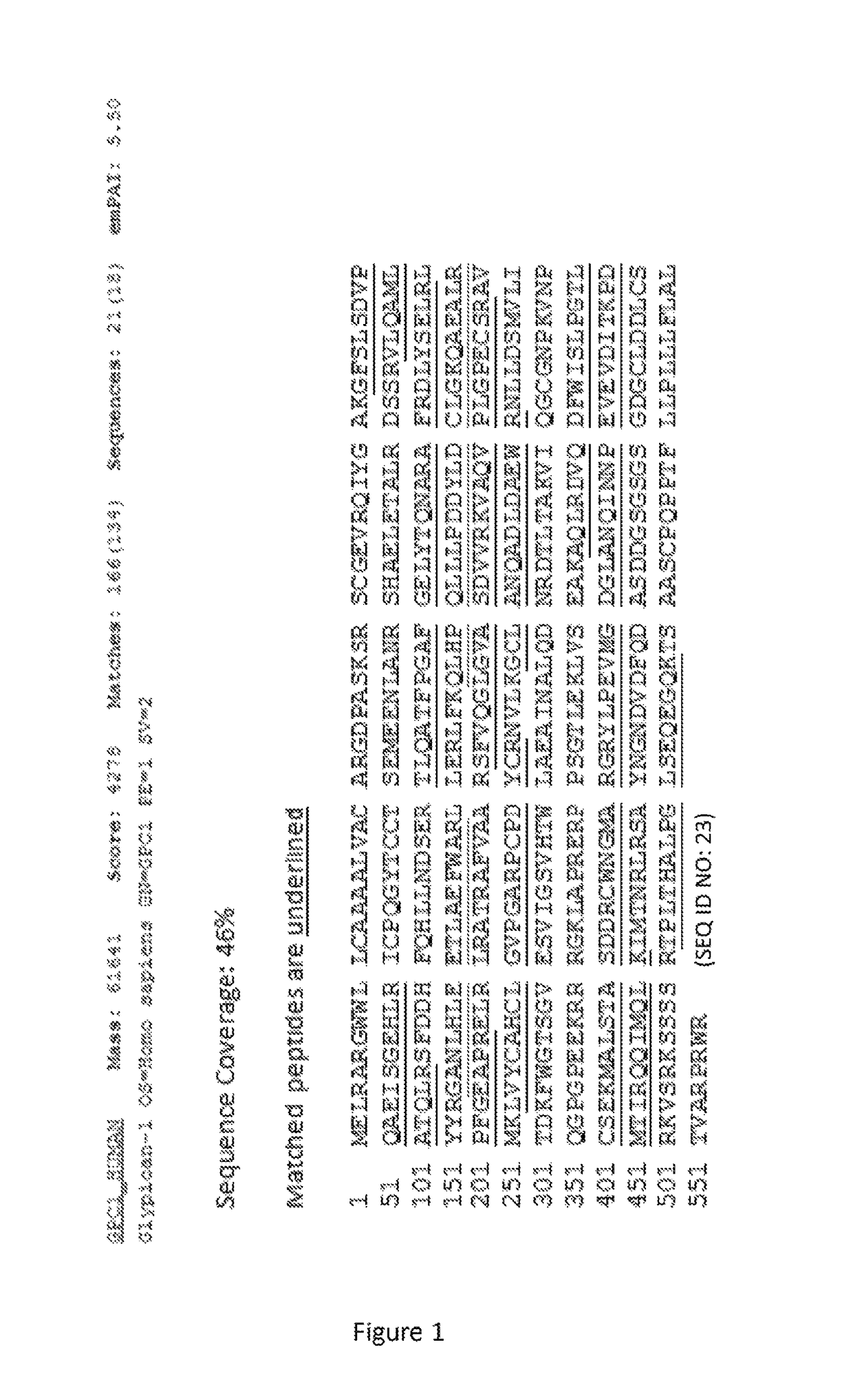
[0141]The mass spectrometer identified glypican-1 with a peptide score of 4278 and sequence coverage of 46% including 18 distinct sequences (FIG. 1).
example 3
tigen Immunoprecipitation, Size Exclusion Chromatography and Mass Spectrometry
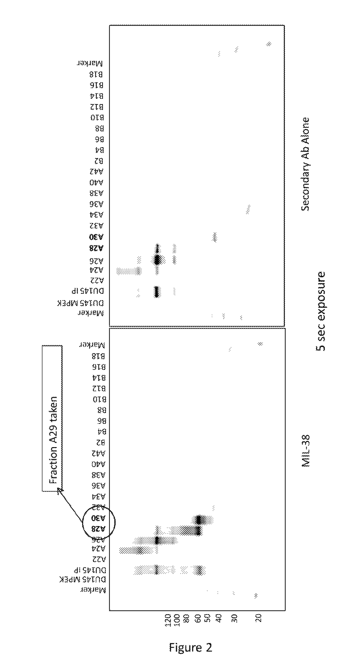
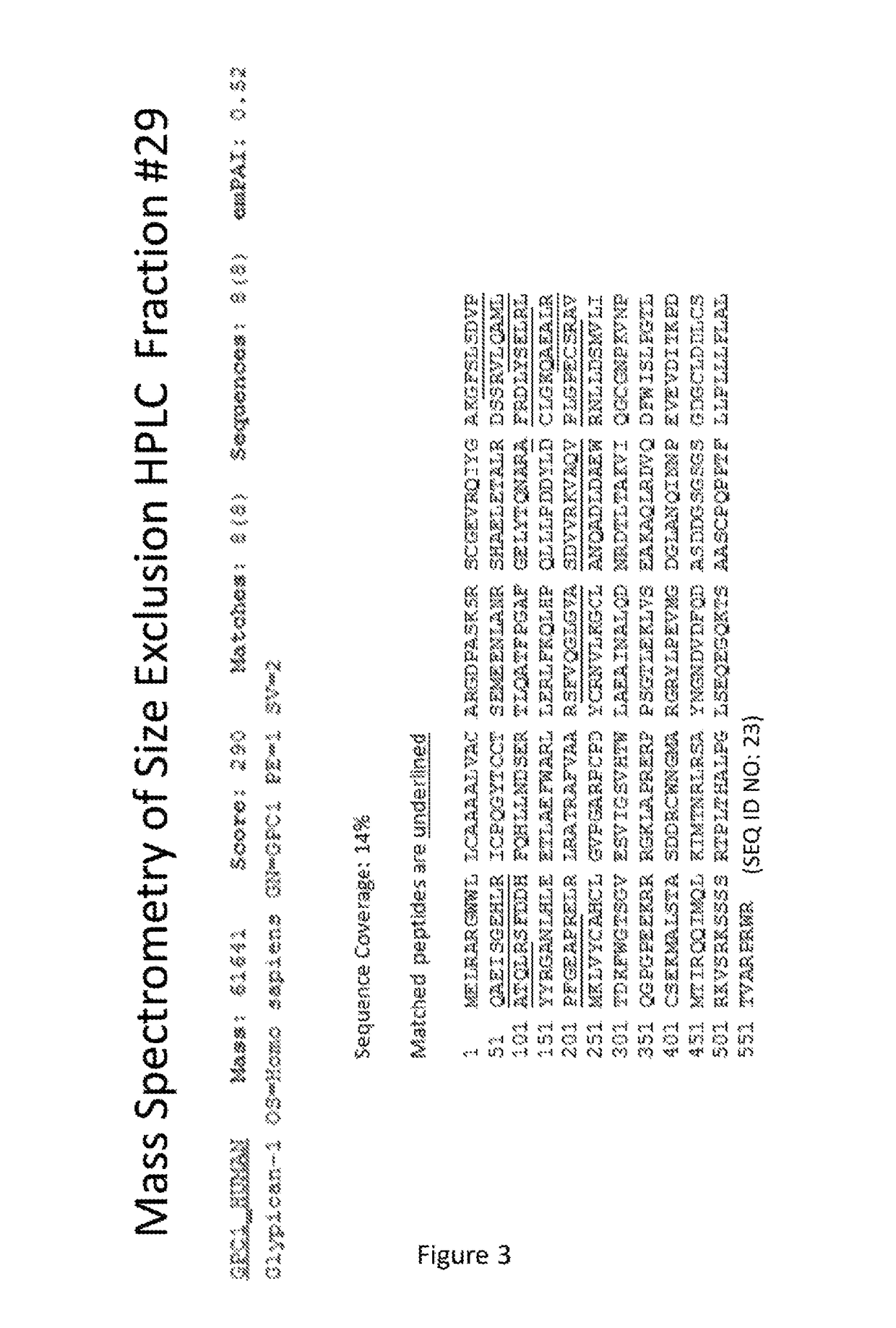
[0142]DU-145 prostate cancer cells were processed through a membrane protein extraction kit (MPEK). Membrane extracts were immunoprecipitated with MIL-38 cross-linked to magnetic beads. Following extensive washing, the immunoprecipitate was eluted in TBS (tris buffered saline) containing 2% SDS. The eluate was subjected to size exclusion chromatography (SEC) and every second fraction was acetone precipitated, resuspended in sample loading buffer and used for MIL-38 western blots. Fractions 28 and 30 contained high amounts of MIL-38 antigen (FIG. 2), indicating that fraction 29 would also contain high concentrations of the MIL-38 antigen. Fraction 29 was subjected to mass spec analysis and glypican-1 was identified with sequence coverage of 14% (FIG. 3). This further confirmed that the antigen for the MIL-38 antibody was glypican-1.
PUM
| Property | Measurement | Unit |
|---|---|---|
| mass spec analysis | aaaaa | aaaaa |
| mass spec analysis | aaaaa | aaaaa |
| pH | aaaaa | aaaaa |
Abstract
Description
Claims
Application Information
 - R&D
- Intellectual Property
- Life Sciences
- Materials
- Tech Scout
- Unparalleled Data Quality
- Higher Quality Content
- 60% Fewer Hallucinations
Browse by: Latest US Patents, China's latest patents, Technical Efficacy Thesaurus, Application Domain, Technology Topic, Popular Technical Reports.
© 2025 PatSnap. All rights reserved.Legal|Privacy policy|Modern Slavery Act Transparency Statement|Sitemap|About US| Contact US: help@patsnap.com


