Colorimetric detection of nucleic acid amplification
- Summary
- Abstract
- Description
- Claims
- Application Information
AI Technical Summary
Benefits of technology
Problems solved by technology
Method used
Image
Examples
example 1
Colorimetric Detection of a Nucleic Acid Amplification Reaction Product
[0079]In an assay for colorimetric detection of a nucleic acid amplification reaction product, the following reagents were mixed together to produce a 2× reagent mix:[0080]Magnesium Sulphate (Sigma Aldrich) at 16 mM[0081]Ammonium Sulphate (Sigma Aldrich) at 20 mM[0082]Potassium Chloride (Sigma Aldrich) at 20 mM[0083]Sodium hydroxide (Sigma Aldrich) at a concentration that sets the starting pH of the reagent mix to 8.8 pH
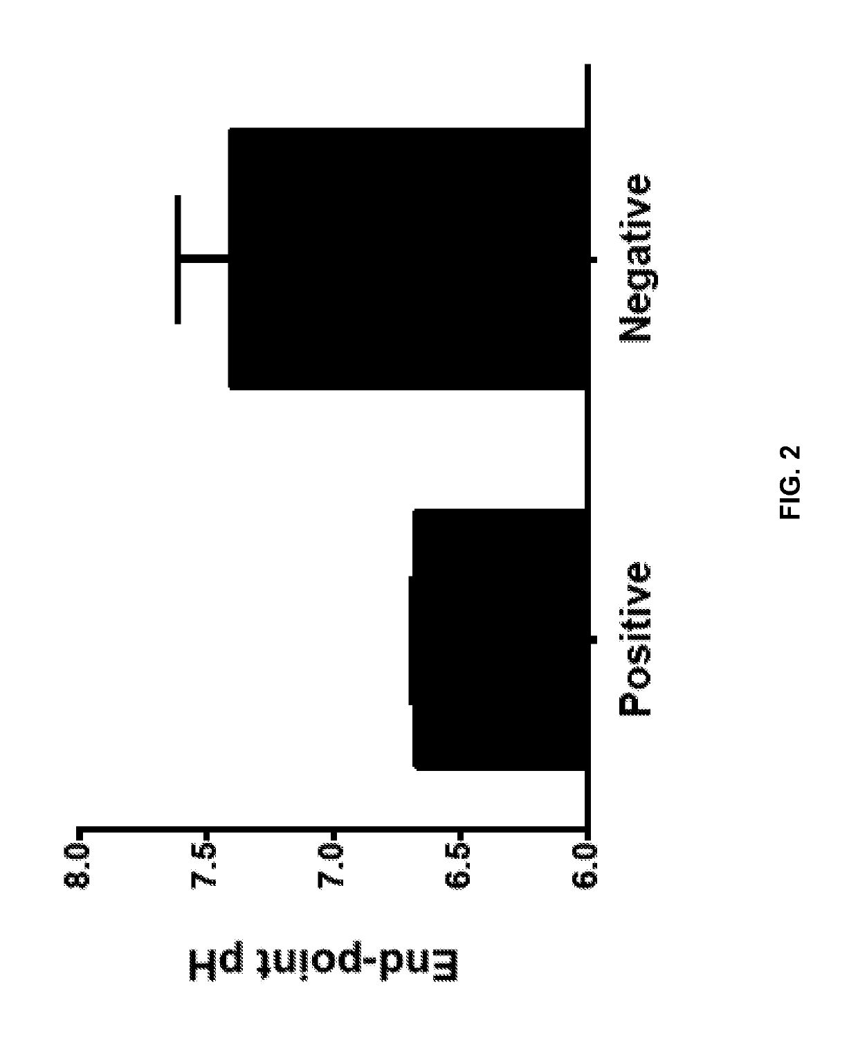
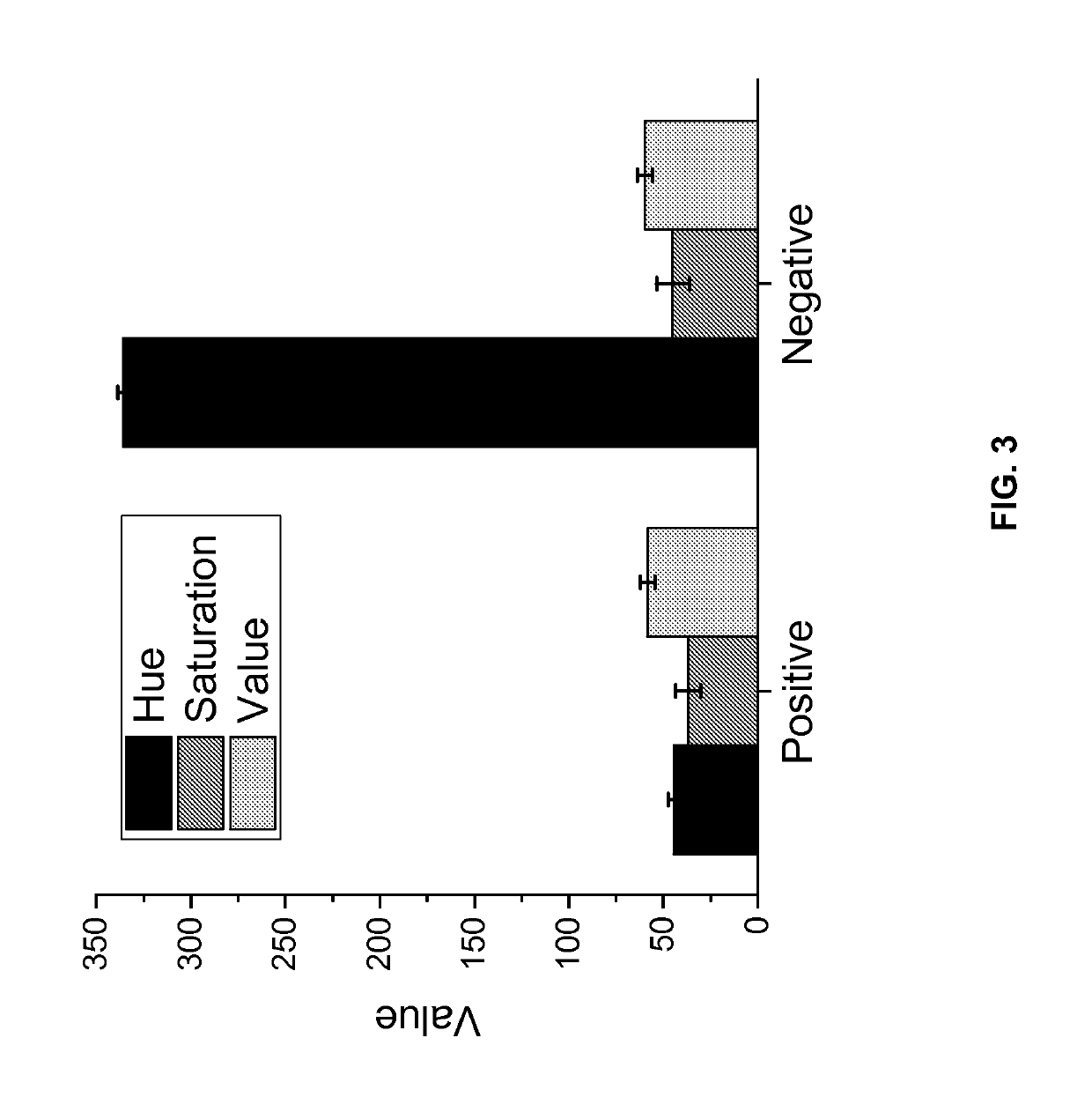
[0084]The reagent mix was adjusted to an initial pH of 8.8 to enable efficient initial polymerization. The reagent mix was autoclaved for 1 hour for sterilization. The following ingredients were then added (in a sterile form) to the reagent mix to generate the reaction mix:[0085]Tween20 (Sigma Aldrich) at 0.1% (v / v)[0086]dNTPs (NEB) at 1.4 mM each[0087]Phenol Red (Sigma Aldrich) at 50 μM[0088]Bst polymerase (NEB) at 0.8 Unit per microliter (the enzyme storage buffer contributing 1 mM Tris buffer, ...
example 2
Detection of LAMP Amplification Using a Visual Halochromic Agent
[0093]LAMP reactions were performed with a reaction mix comprising of: 10 mM (NH4)2SO4, 15 mM KCl, 0.1 mM EDTA, 0.1 mM DTT, 0.01% Triton X-100 (v / v), 5% Glycerol, 8 mM MgSO4, 1.4 mM each dNTPs, 0.1% v / v Tween-20, 0.8 M Betaine. Three primer pairs, specific to different targets, were added to a final concentration of 1.6 μM each for FIP / BIP, 0.2 μM each for F3 / B3, 0.4 μM each for LoopB / F. The final reaction volume is 10 μL, and was held at 63° C. for different incubation times.
[0094]In FIG. 5, the final Tris buffer concentration of the reaction mix was varied from 0.34 mM to 19 mM (by varying amount of Tris buffer formulated to pH 8.8). Reactions were performed with primers for lambda phage DNA, 5 ng of lambda DNA (New England Biolabs), 0.8 U / μl Bst 2.0 DNA polymerase (New England Biolabs) and 0.2 mM Neutral Red (Sigma Aldrich). The reaction tubes were then imaged and the Normalized Hue value was calculated for the color...
example 3
Visual Detection of LAMP Amplification in Sub-Millimeter Path Lengths
[0100]LAMP reactions were performed as in Example 1 with 1.3 mM final Tris buffer concentration (buffer formulated to pH 8.8), 0.8 U / μl of Bst 2.0 DNA Polymerase, 5 ng lambda DNA template and 0.2 mM Neutral Red or 160 μM Bromothymol Blue. Both the positive and the no-template negative reactions were added after amplification to flow chambers with varying channel depths (FIG. 9A for Neutral Red and FIG. 9B for Bromothymol Blue). These flow chambers were machined in acrylic with channel depths ranging from 50 μm to 400 μm. High contrast color difference (above the visual detection threshold; dotted line) between the positive and the negative reactions was observed for channel depths of 50 μm and above. This demonstrates that this visual detection chemistry is amenable for use in reaction chambers with sub-milimeter path lengths (depths) and above. Such reaction chambers can be used to reduce the amount of reagents us...
PUM
| Property | Measurement | Unit |
|---|---|---|
| pH | aaaaa | aaaaa |
| pH | aaaaa | aaaaa |
| pH | aaaaa | aaaaa |
Abstract
Description
Claims
Application Information
 - R&D
- Intellectual Property
- Life Sciences
- Materials
- Tech Scout
- Unparalleled Data Quality
- Higher Quality Content
- 60% Fewer Hallucinations
Browse by: Latest US Patents, China's latest patents, Technical Efficacy Thesaurus, Application Domain, Technology Topic, Popular Technical Reports.
© 2025 PatSnap. All rights reserved.Legal|Privacy policy|Modern Slavery Act Transparency Statement|Sitemap|About US| Contact US: help@patsnap.com


