


Quick Research
Generate reliable direction feasibility study reports for your R&D in just a few steps.

Technical Q&A
Discover and master advanced knowledge NOW. Basics, ideas, possibilities, all at once.

Find Solutions
As an expert in R&D theories, this can generate solutions to your technical problems instantly.

Evaluate Feasibility
Analyze your overall solution with one click, know your potential R&D risks in advance.

Monitor Landscape
Get weekly tech updates, stay abreast of the latest tech innovations and key insights.
Heterocyclic molecules for biomedical imaging and therapeutic applications
a biomedical imaging and therapeutic application technology, applied in the field of heterocyclic molecules for biomedical imaging and therapeutic applications, can solve the problems of limited utility of pib to diagnose and monitor the progression of disease, limit the sensitivity of pet imaging in a prodromal, and limit the diagnostic potential of existing imaging agents to segregate patients, so as to achieve enhanced specificity and enhanced sensitivity
- Summary
- Abstract
- Description
- Claims
- Application Information
AI Technical Summary
Benefits of technology
Problems solved by technology
Method used
Image

Examples
example 1
[0383]This example illustrates an Aβ targeted probe of the present teachings.
[0384]Utilizing the agent F-AI-182, a heterocyclic molecule was synthesized via multiple steps, purified via chromatography, crystallized in methylene chloride and pentane mixture, and the single crystal structure was determined. F-AI-182 was further characterized via standard analytical tools, including 1H NMR, proton-decoupled 13C-NMR, 19F NMR, high resolution mass spectroscopy (HRMS), and analyzed for uniformity using HPLC (Waters) equipped with a dual λ detector (2487) set to 280 and 364 nm on a semi-preparative C-18 column (Vydac).
example 2
[0385]This example illustrates 18F-AI-182 synthesis and testing.
[0386]For bioassays described in following sections, 18F-AI-182 was synthesized via standard nucleophilic substitution, employing 2,2,2-kryptofix / 18F and AI-182-tosylate analog, purified on a C-18 (Vydac) column employing a gradient eluent mixture of ethanol and water, using radio-HPLC system equipped with a radiodetector (Bioscans). The fraction at Rt=15 min was collected, concentrated, and resuspended in PBS to 5% ethanol for all radiotracer bioassays. Furthermore, 18F-AI-182 was also characterized by spiking with an analytically characterized sample of an unlabeled F-AI-182 counterpart, prior to injection on the radio-HPLC.
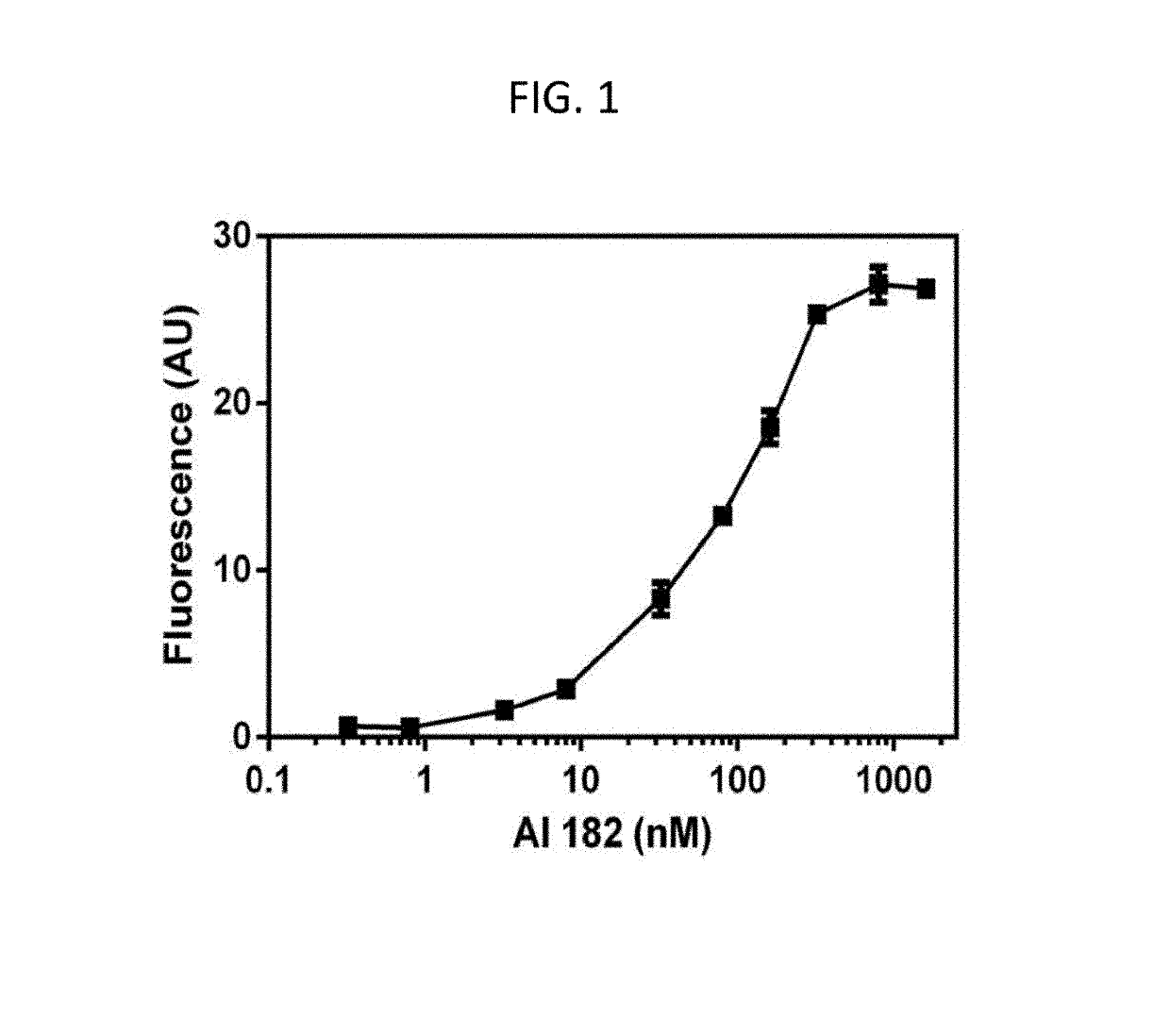
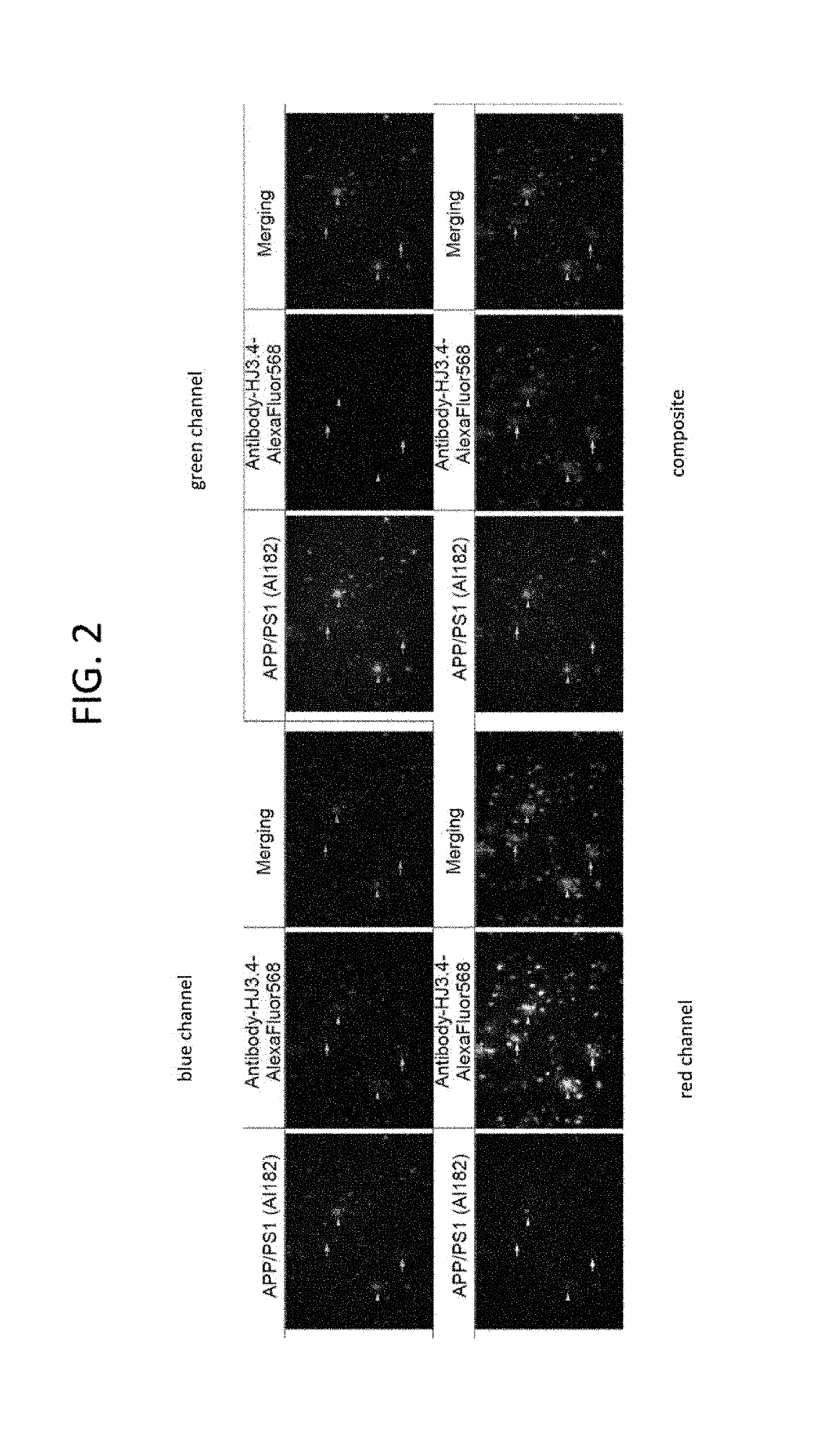
[0387]The agent F-AI-182 shows concentration dependent and saturable binding (binding constant, 59±7 nM; FIG. 1) to preformed Aβ1-42 fibrils, stains both fibrillar and diffuse plaques ex vivo in the hippocampus and cortical region of brain sections in APPsw+ / − / PS1 mice (FIG. 2) and human tissues (F...
example 3
[0389]This example illustrates ex vive staining studies.
[0390]FIG. 2 illustrates ex vivo staining studies were performed on brain sections (50 μm) of an APPsw+ / − / PS1 mouse (24 months old) and a control WT mouse (BL / 6; 24 months old) using well-established procedures. Briefly, tissue sections were immunostained with mouse monoclonal antibody and visualized by donkey-anti-mouse HJ3.4 Aβ monoclonal antibody-Alexa 568 as a positive control. Brain sections of APPsw+ / −PS1+ / −mice showed abundant staining of Aβ compared with minimal levels in WT mouse (FIG. 2). Using F-AI-182 (100 nM, 60 min), abundant staining of fibrillar and diffuse plaques in the hippocampus and cortical regions of brain sections in APPsw+ / −PS1+ / −mice was observed—arrows indicate labeling of Aβ plaques (arrows, diffuse; arrow head, fibrillar). By comparison, no staining in WT mice was seen either with F-AI-182 or the antibody indicating the targeting specificity of F-AI-182. The slides were analyzed on a Zeiss LSM 5 PAS...
PUM
| Property | Measurement | Unit |
|---|---|---|
| Capacitance | aaaaa | aaaaa |
| Pharmaceutically acceptable | aaaaa | aaaaa |
Abstract
Description
Claims
Application Information
 - R&D Engineer
- R&D Manager
- IP Professional
- Industry Leading Data Capabilities
- Powerful AI technology
- Patent DNA Extraction
Browse by: Latest US Patents, China's latest patents, Technical Efficacy Thesaurus, Application Domain, Technology Topic, Popular Technical Reports.
© 2024 PatSnap. All rights reserved.Legal|Privacy policy|Modern Slavery Act Transparency Statement|Sitemap|About US| Contact US: help@patsnap.com