
Methods and system for atrial fibrillation ablation using medical images based cardiac mapping with 3-dimentional (3D) tagging with optional esophageal temperature monitoring

a technology of cardiac mapping and atrial fibrillation, applied in the field of atrial fibrillation ablations, can solve the problems of increased morbidity and mortality, general decrease in quality of life of those afflicted with atrial fibrillation, and atrial fibrillation, so as to minimize the esophageal temperature related injury and eliminate/minimize the effect of esophageal injury during ablation

Active Publication Date: 2019-09-17
AMERICAN MEDICAL TECH LLC
View PDF15 Cites 3 Cited by
  • Summary
  • Abstract
  • Description
  • Claims
  • Application Information

AI Technical Summary

Benefits of technology

The patent describes a system for ablating tissue in the heart to treat atrial fibrillation. The system uses a combination of fluoroscopy and other medical images to guide the placement of balloon catheters for ablation procedures. The system also includes a temperature control system to monitor and regulate the temperature of the ablation catheter. The technical effects of the patent include improved placement of the balloon catheter for atrial fibrillation ablations and increased safety during the procedure.

Problems solved by technology

Atrial fibrillation is associated with increased morbidity and mortality, and in particular, a general decrease in quality of life for those afflicted with atrial fibrillation.
AF can also cause tachycardia mediated cardiomyopathy or worsening of pre-existing heart failure.
Patients are at an increased risk of stroke unless they are treated adequately with anticoagulants.
Anticoagulant treatment however, increases the patient's risk of bleeding, which carries with it is own set of dangers.
Medications currently available for treating atrial fibrillation have proven to be only moderately effective in decreasing the incidence of recurrent atrial fibrillation, and these medications do not decrease the patient's risk of having a stroke.
Moreover, the AV nodal artery passes near the mouth of the coronary sinus, the ablation may conceivably damage this vessel.
RF ablation may be painful to the patient under conscious sedation through direct stimulation of cardiac sensory nerves or pericardial or collateral Whisler visceral irritation, particularly when ablating your thin-walled or venous structures such as posterior left atrium, coronary sinus, or posterior cable tricuspid Isthmus.
This first select procedures associated with substantial patient discomfort, the use of cryoablation may theoretically result in lower anesthetic and analgesic requirements.

Method used


Image

  • Methods and system for atrial fibrillation ablation using medical images based cardiac mapping with 3-dimentional (3D) tagging with optional esophageal temperature monitoring
  • Methods and system for atrial fibrillation ablation using medical images based cardiac mapping with 3-dimentional (3D) tagging with optional esophageal temperature monitoring
  • Methods and system for atrial fibrillation ablation using medical images based cardiac mapping with 3-dimentional (3D) tagging with optional esophageal temperature monitoring

Examples


Embodiment Construction

[0226]The following description is of the best mode presently contemplated for carrying out the disclosure. This description is not to be taken in a limiting sense, but is made merely for the purpose of describing the general principles of the disclosure.

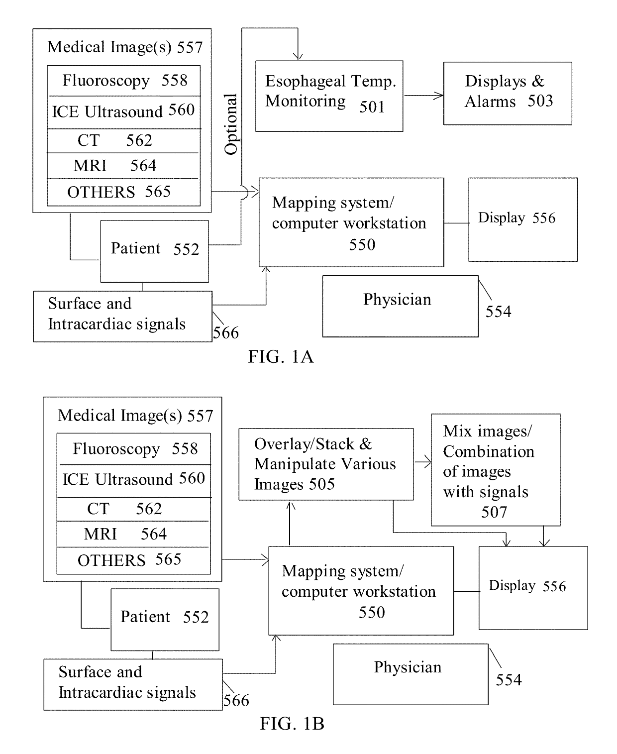
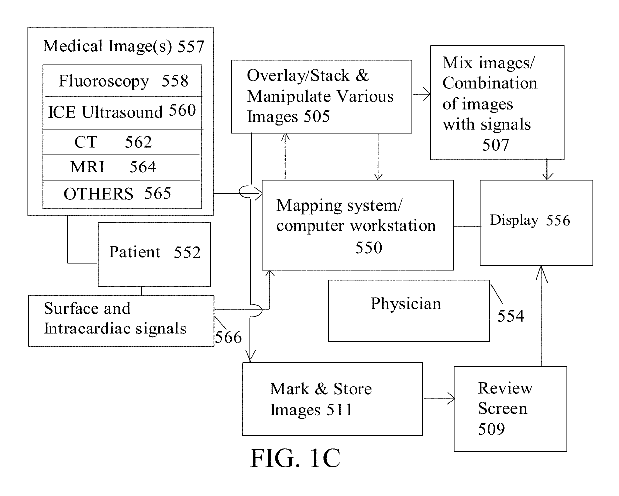
[0227]In the methods and system of this disclosure, medical images based cardiac mapping / electrophysiology tools is disclosed for cardiac ablations for arrhythmias. The methods and system of this disclosure can be employed with a methodology for monitoring esophageal temperature. In another embodiment, the mapping system and mapping methodology can also be used without the use of temperature monitoring.

Definitions:

[0228]In this disclosure, cardiac mapping is defined and used as a guide for physicians to utilize during the cardiac ablation procedure, which includes making maps, guiding physicians to the optimal placement of the catheters, utilizing various types of medical images or a combination of medical images. Cardiac mapping al...


PUM


Abstract

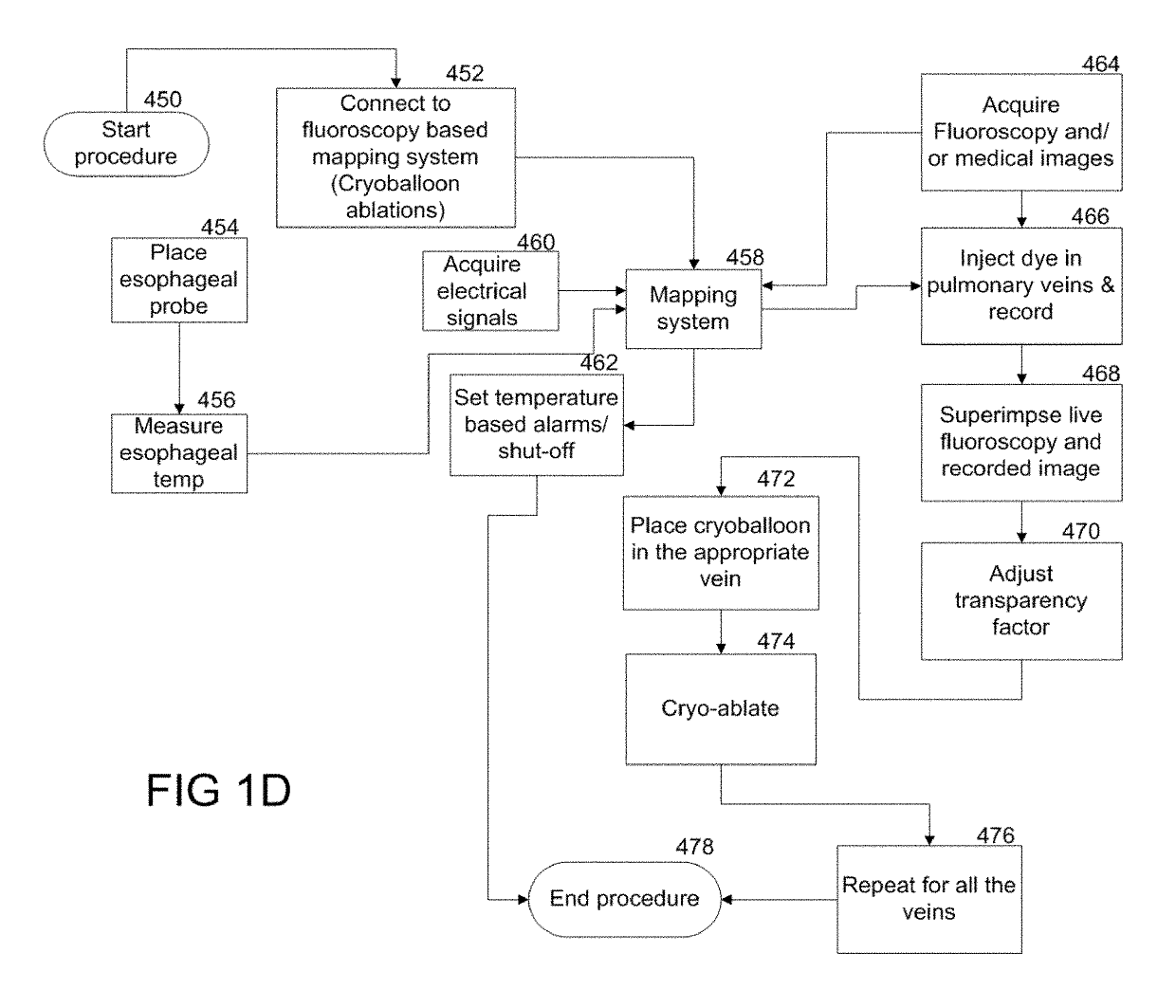
Methods and system for atrial fibrillation ablations utilizing cardiac mapping based on medical image(s). The method and system also adapted for any balloon based catheters including cryoballoon catheter, laser balloon catheter, or “hot ballooncatheter, as well as circular catheters. The methods and system includes overlaying of two or more images on top of each other (where the images are of various types) and aligning the images, where the transparency between the images can be adjusted as an aid for optimal placement of the balloon based catheters or circular catheters. 3D volumetricTags and markers are also placed on 3D images such as CT and MRI images which are indicative of where the tissue has been ablated. The method and system also comprises the ability to monitor esophageal temperature, and to activate alarms and / or energy delivery interrupt based on pre-determined esophageal temperature parameters.

Description

[0001]This application is a Continuation-in-part (CIP) of U.S. application Ser. No. 15 / 209,265 filed Jul. 13, 2016. This application also claims priority of the following provisional patent applications.[0002]METHOD AND SYSTEM FOR CARDIAC MAPPING UTILIZING VARIOUS MEDICAL IMAGING MODALITIES OR COMBINATION THEREOF FOR CARDIAC ABLATIONS—Application number 62 / 536,011 filed Jul. 24, 2017[0003]FLUOROSCOPY AND CARDIAC IMAGE(S) BASED MAPPING SYSTEM FOR GUIDING CRYOBALLOON ABLATIONS FOR ATRIAL FIBRILLATION—Application number 62 / 343,745 filed May 31, 2016[0004]FLUOROSCOPY AND CARDIAC IMAGE(S) BASED MAPPING SYSTEM FOR GUIDING CRYOBALLOON ABLATIONS FOR ATRIAL FIBRILLATION WITH AUTOMATIC FLUOROSCOPIC RECORDING MECHANISM—Application number 62 / 346,539 filed Jun. 6, 2016[0005]MAPPING METHOD AND SYSTEM FOR CRYOBALLOON ABLATION FOR ATRIAL FIBRILLATION—Application number 62 / 429,691 filed Dec. 2, 2016FIELD OF DISCLOSURE[0006]The present disclosure relates to atrial fibrillation ablations, more specifi...

Claims


Application Information

Patent Timeline
Patent Type & Authority Patents(United States)
IPC IPC(8): A61B5/00A61M25/10A61B8/12A61B18/14A61B18/28A61B18/00A61B8/00A61B5/046A61B5/042A61B5/01A61B90/00A61B5/361
CPCA61B18/1492A61B8/12A61B18/28A61M25/1018A61B5/0035A61B5/0044A61B5/046A61B2018/00702A61B5/042A61B90/36A61B2018/00791A61B8/445A61B2018/00577A61B2018/00351A61B5/015A61B8/0883A61B6/485A61B6/481A61B6/5247A61B5/283A61B5/361
Inventor BOVEJA, BIRINDER R.WIDHANY, ANGELY
Owner AMERICAN MEDICAL TECH LLC
Learn More