

Imaging biomarkers for the diagnosis and prognosis of back pain and related conditions
a biomarker and back pain technology, applied in the field of imaging biomarkers, can solve the problems of not being able to find reliable noninvasive methods to detect painful discs, the link between degeneration and pain is not always present, and the non-availability of non-invasive methods, so as to reduce the avoid fat signal interference, and reduce the effect of field of view excitation
- Summary
- Abstract
- Description
- Claims
- Application Information
AI Technical Summary
Benefits of technology
Problems solved by technology
Method used
Image

Examples
example 1
Introduction
[0042]As indicated above, low back pain (LBP) is a disease with wide prevalence and significant burden. In the adult population, back pain of at least moderate intensity and duration has an annual incidence of 10-15%. It adversely influences the lives of those affected, resulting in suffering and disability, and poses enormous economic burden on individuals and the society. Back pain is associated with natural degenerative process of the intervertebral disc (IVD), which is the largest avascular structure in the human body. The IVD is composed of: (1) the nucleus pulposus (NP), a gel-like structure in the center of the disc rich in proteoglycan [protein core with glycosaminoglycans (GAGs)]; (2) the annulus fibrousus (AF), a collagenous zone around the NP; and (3) the cartilaginous end-plate (EP) on top and bottom (3). GAGs play a critical role in supporting IVD functions including generating hydrostatic pressure to resist loading by binding water. The loss of GAGs is coup...
example 2
Materials and Methods
Phantom
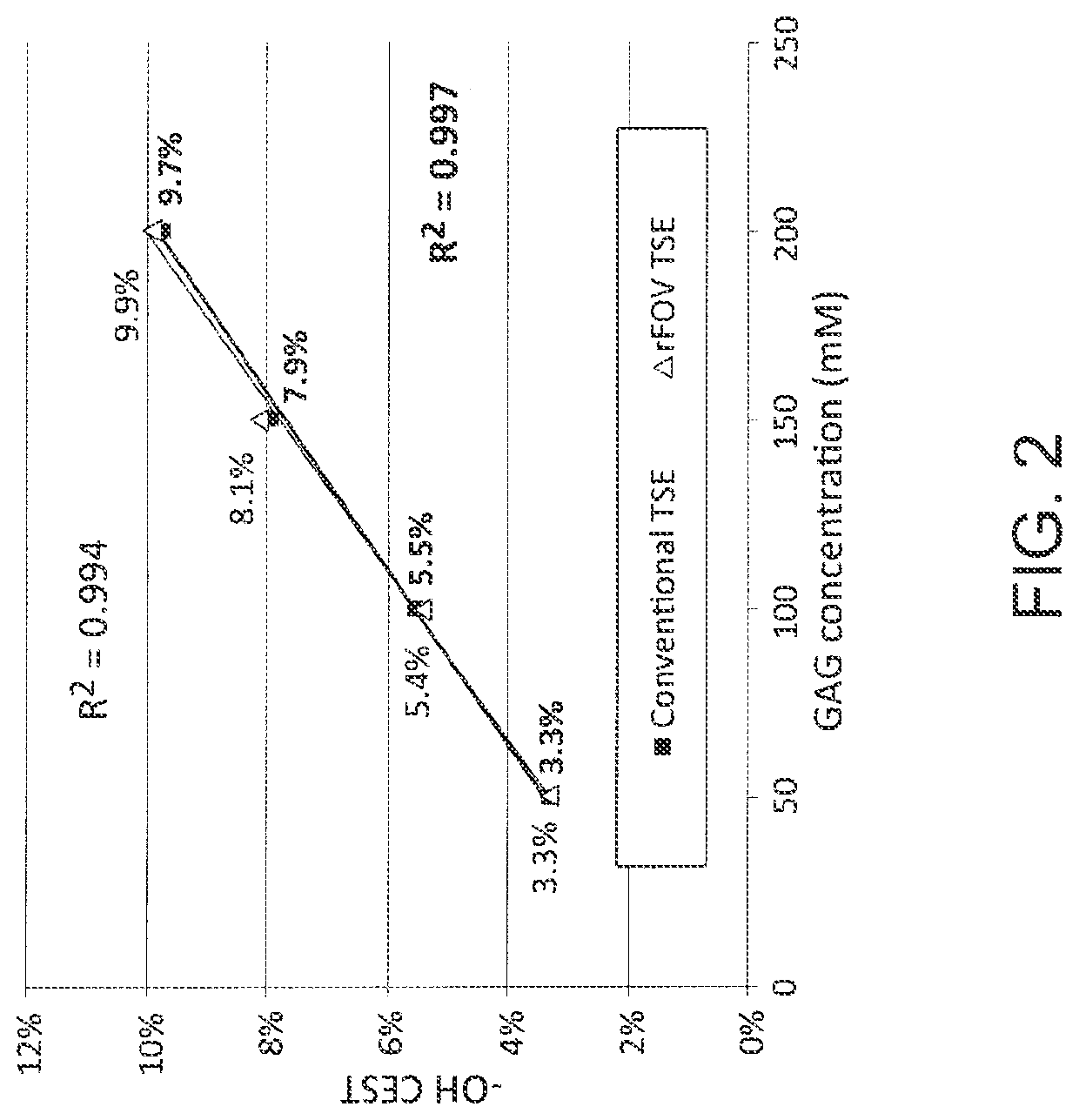
[0047]To verify the relationship between GAG concentration and CEST signal, four samples of GAGs with concentrations of 50, 100, 150, and 200 mM respectively were prepared from chondroitin sulphate A (Aldrich-Sigma, St. Louis, Mo., USA) in a standard solution of phosphate-buffered saline. All pHs were titrated to 7.0. The concentration refers to the number of disaccharide units in GAGs. At the time of imaging the GAG samples were individually placed in a gadolinium-doped water bath to reduce its T1.
Human Subjects
[0048]Nine healthy volunteers (3 female, 6 male; mean age 39.1±11.9) with no symptoms related to the spine and no history of spine disease were recruited.
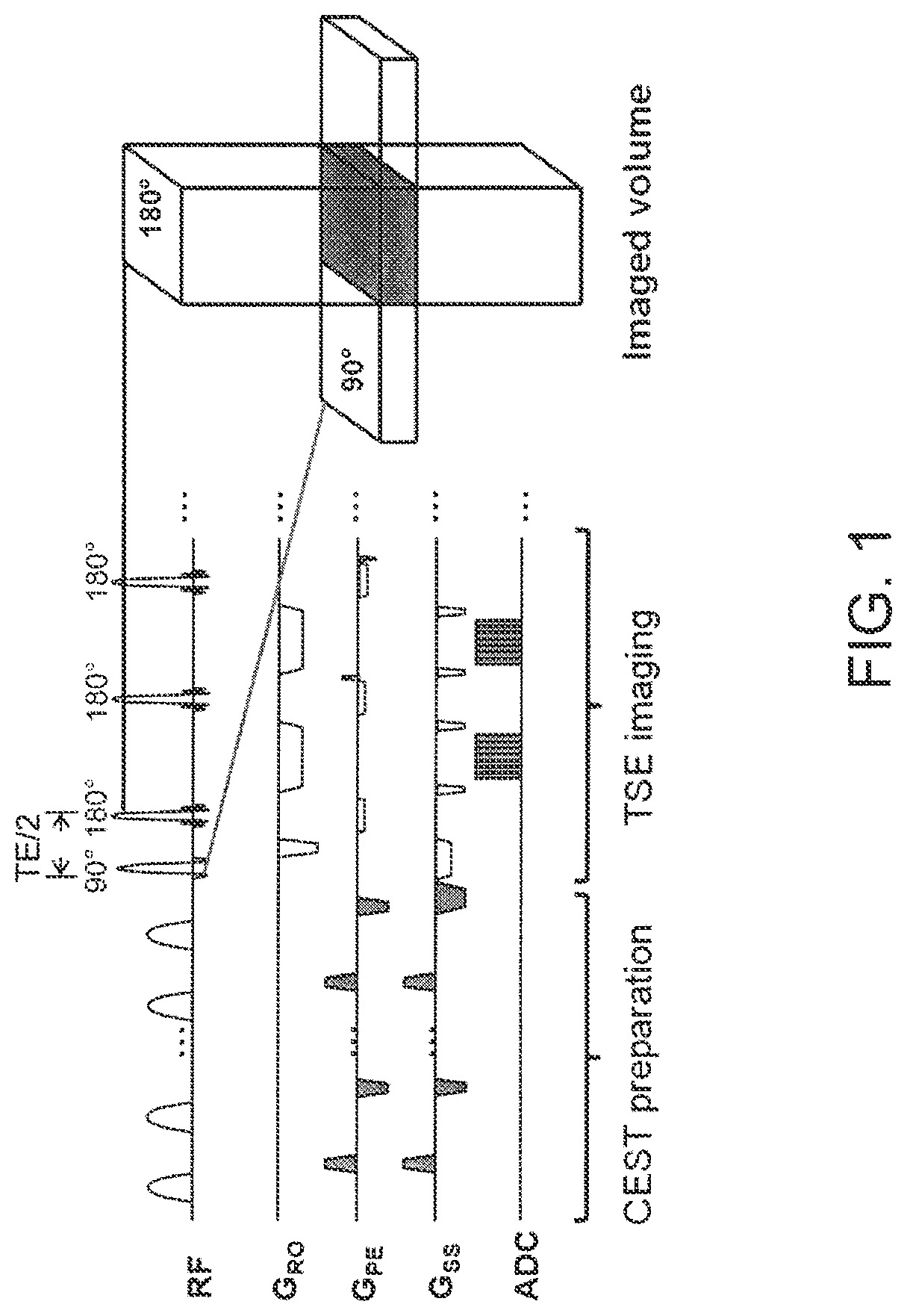
[0049]A 2D rFOV TSE CEST sequence (FIG. 1) was implemented. To achieve rFOV, gradients of the 180° refocusing pulses in TSE were moved from the slice-encoding direction to the phase-encoding direction with their magnitude modified accordingly based on the desired reduced FOV size. A...
example 3
[0054]All CEST images were first normalized by S0 and corrected for B0 inhomogeneity pixel by pixel. The pixel-by-pixel B0 frequency shift was determined in WASSR by fitting a Lorentzian line shape (See Zaiss et al. Quantitative separation of CEST effect from magnetization transfer and spillover effects by Lorentzian-linefit analysis of z-spectra. J. Magn. Reson. 2011; 211(2):149-155, which is incorporated by reference herein in its entirety as though fully set forth) in a 0.8 ppm frequency range which has substantial saturation. Then the curve was spline interpolated to produce the Z-spectrum (CEST vs. offset-frequency curve). Data at ±4.5 ppm were excluded from subsequent analysis. Magnetization transfer asymmetry ratio (MTRasym) for each pixel was calculated as:
[0055]MTRasym(Δω)=S(-Δω)-S(Δω)S0,[1]
where Δω is the frequency offset of the saturation pulse, S(±Δω) is the CEST signal intensity at the frequency offset +Δω and −Δω respectively.
[0056]For each phantom, o...
PUM
 Abstract
Description
Claims
Application Information