Antibodies for treatment and diagnosis of inflammatory bowel disease

a technology for inflammatory bowel disease and antibodies, applied in the field of disease diagnosis and treatment, can solve the problems of not being able to fully explain the presence or role of iiics in inflammatory bowel disease, not being suitable for targeting applications, etc., and achieves the effect of facilitating production and purification and facilitating clinical-grade material

Active Publication Date: 2020-05-12
PHILOGEN SRL LLC
View PDF1 Cites 1 Cited by
  • Summary
  • Abstract
  • Description
  • Claims
  • Application Information

AI Technical Summary

Benefits of technology

[0043]An antibody molecule of the present invention may be conjugated to a molecule to provide a conjugate. The choice of molecule conjugated to the antibody molecule will depend on the intended application of the conjugate. For example, where the conjugate is intended for the treatment of an inflammatory disorder or autoimmune disease, the conjugate may comprise an antibody molecule of the invention, or for use in the invention, and an immunosuppressive or anti-inflammatory agent, such as a cytokine. Where the conjugate is intended for use in imaging, detecting, or diagnosing a disease or disorder, the conjugate may comprise an antibody molecule of the invention, or for use in the invention, and a detectable label, such as a radioisotope, e.g. a non-therapeutic radioisotope. Depending on the molecule conjugated to the antibody molecule, the conjugate may be or may comprise a single chain protein. When the conjugate is a single chain protein, the entire protein can be expressed as a single polypeptide or fusion protein. In this case, the molecule may be conjugated to the antibody molecule by means of a peptide linker. Fusion proteins have the advantage of being easier to produce and purify since they consist of one single species. This facilitates production of clinical-grade material. Alternatively, the molecule may be conjugated to the antibody molecule by means of a cleavable linker.

Problems solved by technology

However, since the antibody requires both the peptide backbone and the carbohydrate moiety to recognize the epitope, it is not suitable for targeting application especially when cross-reactivity between species is needed.
However, to our knowledge there is no report on the presence or role of IIICS in Inflammatory Bowel Disease.
However, the role of the A1 and of the D domain of Tenascin-C has not been elucidated in full and is not clear whether they can be used as a target for the pharmacodelivery of agents to treat or diagnose IBD.

Method used


Image

  • Antibodies for treatment and diagnosis of inflammatory bowel disease
  • Antibodies for treatment and diagnosis of inflammatory bowel disease
  • Antibodies for treatment and diagnosis of inflammatory bowel disease

Examples


example 1

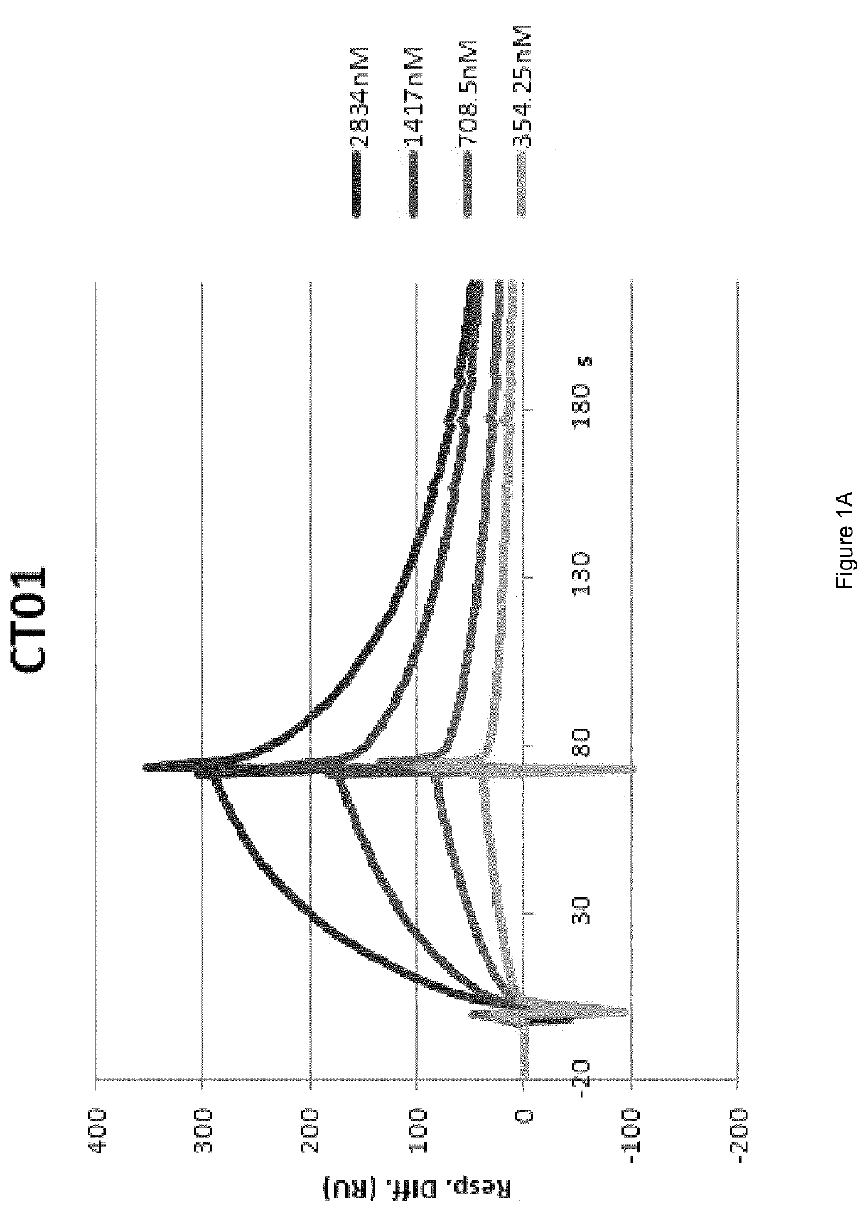
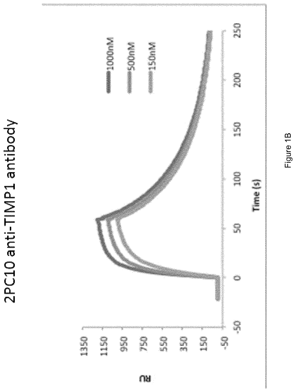
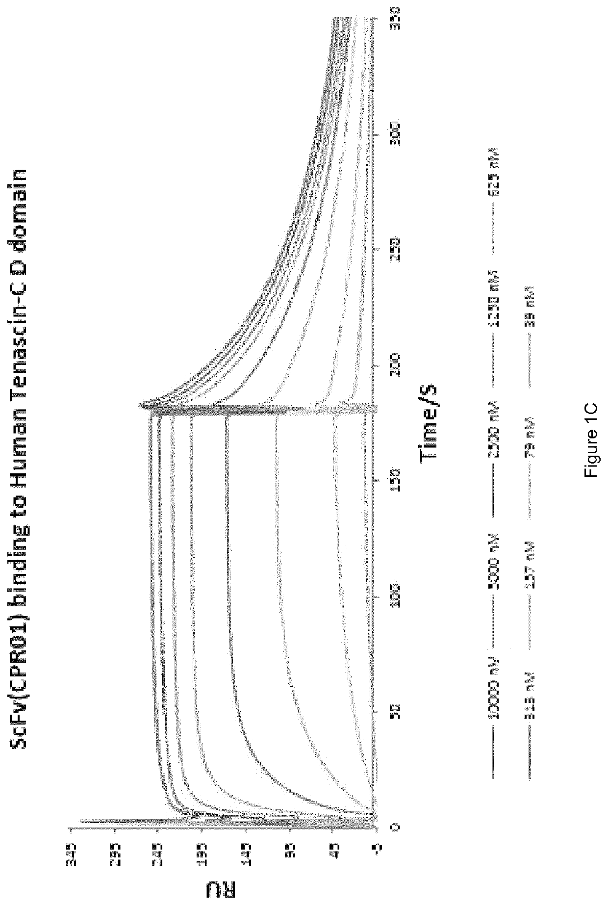
Preparation and Characterisation of the Novel Antibodies CT01 against Lysozyme, FF01 and FF02 against Neutrophil Elastase, 2PC10 against TIMP-1, CPR01 and CPR01.1 against Domain D of Tenascin C

[0130]The antibodies against human lysozyme, human neutrophil elastase, human TIMP-1 and domain D of human tenascin C, antibodies CT01, FF01 and FF02, 2PC10, CPR01 and CPR01.1, were isolated in single-chain Fv (scFv) configuration from phage display libraries, which included the libraries described in PCT / EP2009 / 006487, Weber et al. (PLoS One, 2014, 9 (6) doi: 10 / 1361) and Silacci et al. (Protein Engineering Design & Selection, 2006, 19, 471-478), according to the screening technique described by Silacci et al. (Protein Engineering Design & Selection, 2006, 19, 471-478) using lysozyme, neutrophil elastase, TIMP-1, and domain D of tenascin C, respectively, as the screening antigen.

[0131]Antibodies in SIP format were prepared by fusing the VL domain sequence of the antibody in scFv format to the...

example 2

Immunofluorescence Staining of Sections from Ulcerative Colitis Patients

[0163]Freshly frozen biopsy samples of Ulcerative Colitis were stained according to published methods (S. Pfaffen. Eur J Nucl Med Mol Imaging 2010). In brief, purified biotinylated antibodies in SIP (KSF) or IgG (SW01, L19, F16, CH01, G11) format were added at the final concentration of 2pg / ml to the sections. Detection of the primary antibody was performed with streptavidine-Alexa-488 antibody (Invitrogen).

[0164]Positive control was performed by staining blood vessels with an antibody (eBioscience, 1:100) specific for CD31, an endothelial cell marker, the signal revealed with goat anti-mouse Alexa-594 (Invitrogen). Further positive control was performed by counterstaining for cell nuclei was performed with DAPI (eBioscience). Sections were mounted with fluorescent mounting medium (DAKO) followed by analysis using an Axioskop2 microscope with a 10× objective (Carl Zeiss A G, Jena, Germany). The results are shown...

example 3

Immunofluorescence Staining of HT29 Cells with the CT01 Antibody

[0165]Coverslips were placed into culture dishes (100 mm×20 mm), and HT29 cells (both human adenocarcinoma cell lines) were then seeded at 100,000 cells / mL in the culture dishes respectively. The culture was incubated until proper confluency was reached. Cells were fixed and permeabilized by ice-cold methanol incubation at −20° C. Cells were then blocked with 10% FBS / 2% BSA. Affinity-purified antibody CT01, in SIP format (final concentration 5 μg / ml) was first incubated with the cells, followed by incubation with a rabbit anti-human IgE antibody (1:500 dilution; DAKO). The antibody was then detected with goat anti-rabbit IgG Alexa 488 (1:1000 dilution; Invitrogen). DAPI was used for nuclei staining. The anti-hen egg Lysozyme SIP antibody (KSF) was used as an isotype-negative control for the staining. The results demonstrate that the anti-human lysozyme antibody CT01 is capable of staining lysozyme in biological samples ...


PUM

PropertyMeasurementUnit
flow rateaaaaaaaaaa
flow rateaaaaaaaaaa
concentrationsaaaaaaaaaa

Abstract

The invention relates to the diagnosis and treatment of diseases, including inflammatory disorders, proliferative disorders and autoimmune diseases. The invention provides, and involves the use of, antibody molecules that bind: i) lysozyme, ii) neutrophil elastase, iii) tissue inhibitor of metalloproteinase-1 (TIMP-1), or iv) the D domain of Tenascin-C. The invention also relates to the use of antibody molecules that bind v) the IIICS isoform of fibronectin, vi) the Extra Domain-B (ED-B) of fibronectin, vii) matrix-metalloproteinase 3 (MMP3), or viii) the A1 domain of tenascin-C in the diagnosis and treatment of inflammatory disorders such as inflammatory bowel disease.

Description

FIELD OF THE INVENTION[0001]The present invention relates to the diagnosis and treatment of diseases, in particular inflammatory disorders and autoimmune diseases, such as inflammatory bowel disease (IBD). In this context, the invention provides, and involves the use of, antibody molecules that bind: i) lysozyme, ii) neutrophil elastase, (iii) tissue inhibitor of metalloproteinase-1 (TIMP-1), or (iv) the D domain of tenascin-C. The invention also relates to the use of antibody molecules that bind (v) the IIICS isoform of fibronectin, (vi) the Extra Domain-B (ED-B) of fibronectin, (vii) matrix-metalloproteinase 3 (MMP3), or (viii) the A1 domain of tenascin-C in the diagnosis and treatment of IBD.BACKGROUND TO THE INVENTION[0002]Most conventional pharmaceuticals currently in use for the treatment of serious disorders such as cancer and inflammatory diseases do not selectively accumulate at the site of disease [Bosslet et al., 58, 1195-1201 Cancer Res. (1998)]. For example, intravenous...

Claims


Application Information

Patent Timeline
Patent Type & Authority Patents(United States)
IPC IPC(8): C07K16/18A61K39/00A61K47/68C07K16/28C07K16/40
CPCC07K16/18A61K47/6813A61K47/6843C07K16/28C07K16/40A61K2039/505C07K2317/528C07K2317/60C07K2317/565C07K2317/56C07K2317/92C07K2317/33C07K2317/21C07K2317/622A61P1/04A61P29/00A61P37/02A61P37/06
Inventor WULHFARD, SARAHVILLA, ALESSANDRAPEMBERTON ROSS, CATHERINEPRETTO, FRANCESCAFLEETWOOD, FILIPPA
Owner PHILOGEN SRL LLC
Why Eureka
Try Eureka