
Shaped epicardial lead and placement system and method

a technology of epicardial leads and shaped leads, applied in the field of epicardial leads, can solve the problems of long hospitalization time, large drawbacks of current epicardial leads, and invasive surgical approaches, and achieve the effect of improving heart beating function

Active Publication Date: 2020-05-26
KOBARA MEDICAL INC
View PDF50 Cites 8 Cited by
  • Summary
  • Abstract
  • Description
  • Claims
  • Application Information

AI Technical Summary

Benefits of technology

The present invention is a device that solves issues with existing therapies for heart rhythm disorders by using a single epicardial lead with multiple zones of electrodes that can be placed over different chambers of the heart. This leads to more comprehensive therapy that includes pacing and synchronization of the chambers to improve heart function. The lead has a feature to prevent it from slipping out of place, and the electrodes are spaced along its length to position each one over a specific chamber of the heart.

Problems solved by technology

However, currently marketed epicardial lead designs have major drawbacks.
The first drawback is that current epicardial leads require a more invasive surgical approach to gain access to the epicardium and to be able to fixate the leads into the heart wall.
This approach is surgical in nature, requires anesthesia and puts the patient on single lung ventilation, all of which result in longer hospitalization, higher costs, higher morbidity and present the risk of other complications.
Because of the nature of the access approach, this technique also limits where the leads can be placed, resulting in non-optimal placement of epicardial leads—e.g., intended left ventricular pacing.
The second drawback to conventional techniques is the securing of the lead in its intended position, which is with sutures in combination with active fixation type anchors.
This involves trauma to the heart wall and, even though the fixation is into the tissue, there are still signal capture issues.
The addition of a drug to the implant complicates the development, regulatory approval, and manufacturing of these leads.
The third drawback of current epicardial leads is that the number of contacts or electrodes is limited and local to attachment to the heart wall.
Also, with only a few electrodes limited to a local region, there is no ability to implement advanced signal interpretation algorithms to improve therapy delivery—e.g., reducing inappropriate shocks for devices and leads with defibrillation.
Their use has been very limited due to these and other drawbacks.
Although subcutaneous ICD devices have good sensing capabilities suitable for the intended ICD therapies such as defibrillation, there are many patients who are not ideal candidates as the ECG (electrocardiogram) criteria does not hold well for their candidacy.
Also, current devices lack pacing capability.
This limits their usefulness because the target patients for these devices have advanced heart conduction system disease even in the absence of heart block that requires pacing for heart rhythm management.
Endovascular based pacing leads have limitations as well.
The current traditionally-placed coronary sinus leads used for endovascular left ventricle (LV) pacing for cardiac resynchronization are limited not just by the presence of coronary sinus branches and their caliber, but also by the challenges associated with implantation.
Accordingly, outcomes are limited due to the limitations for pacing.
Even leadless endovascular pacing devices have severe complication risks, including heart wall perforation and dislodgement.
Finally, there are no left atrial pacing or sensing systems which could enable right atrium to left atrium synchronization.
However, it still requires fixation to the heart wall and therefore has similar drawbacks to current leads.
Here too, fixation of the multiple lead branches and electrodes poses significant challenges.
These epicardial lead examples and their associated implantation techniques are not well developed because they are surgical in nature and, consequently, more invasive.
All the identified challenges have limited the implementation of epicardial leads.

Method used


Image

  • Shaped epicardial lead and placement system and method
  • Shaped epicardial lead and placement system and method
  • Shaped epicardial lead and placement system and method

Examples


Embodiment Construction

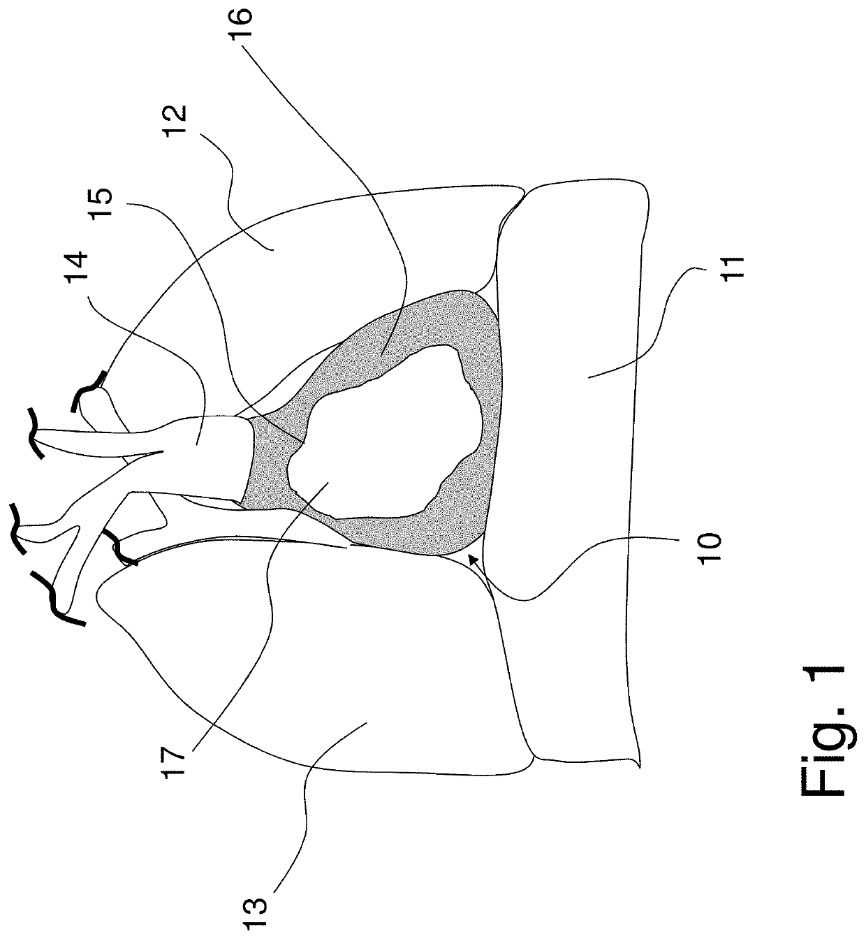
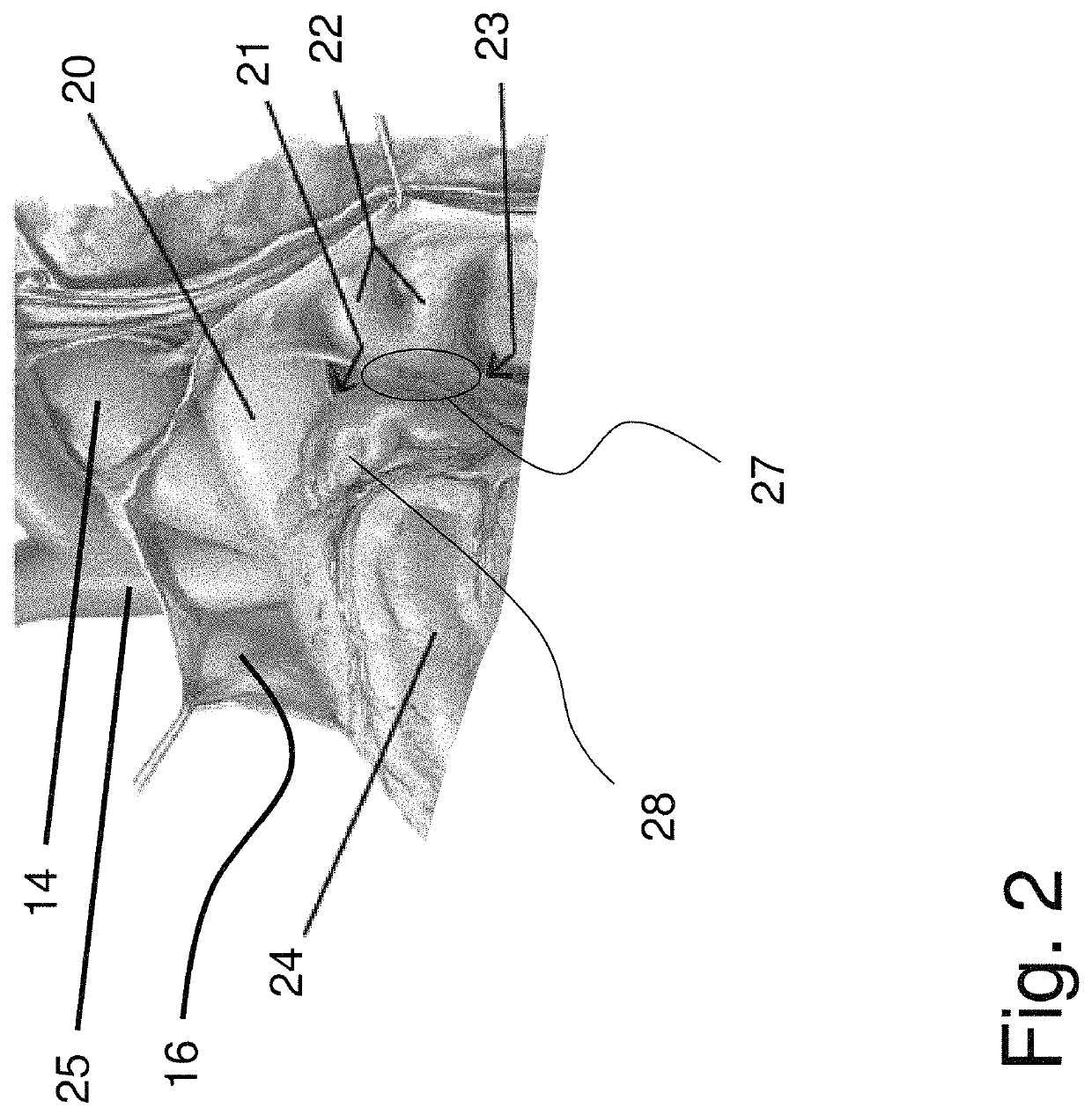
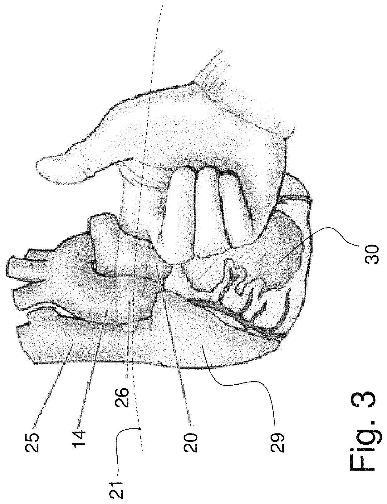
[0045]Exemplary embodiments of the present invention are described in the following, with reference to the drawings. It should be understood that such embodiments are by way of example only and merely illustrative of the many possible embodiments which can represent applications of the principles of the present invention. Various changes and modifications obvious to one skilled in the art to which the present invention pertains are deemed to be within the spirit, scope, and contemplation of the present invention as further defined in the appended claims. The exemplary embodiments of the present invention described herein are not intended to be exhaustive or to limit the present invention to the precise forms disclosed in the following detailed description. Rather, the exemplary embodiments described herein are chosen and described so those skilled in the art can appreciate and understand the principles and practices of the present invention.

[0046]Referring to FIG. 1, an anterior vie...


PUM


Abstract

A cardiac lead system is provided. The lead is placed epicardially through the transverse pericardial sinus with integrated curvatures to prevent the lead from slipping out of the transverse pericardial sinus. Interaction with multiple chambers of the heart is facilitated in a single lead, without anchors that embed into the heart wall. Multiple electrodes can be grouped over each targeted heart area to ensure adequate electrical contact.

Description

PRIORITY[0001]This Application is a continuation application of PCT Patent Application No. PCT / US2017 / 025662, filed Apr. 1, 2017, which claims priority to and the benefit of U.S. Provisional Patent Application No. 62 / 316,871, filed Apr. 1, 2016, and U.S. Provisional Patent Application No. 62 / 409,857, filed Oct. 18, 2016; with each of the above-referenced applications and disclosures incorporated fully herein by reference.FIELD OF THE INVENTION[0002]The present invention relates generally to devices that continuously monitor a patient's cardiac rhythm and deliver electrical pulses to the heart if and as needed. More specifically, the present invention relates to epicardial leads that monitor electrical signals from the epicardial surface of the heart, that delivers electrical energy, or both.BACKGROUND OF THE INVENTION[0003]In the field of cardiac medicine, minimally invasive therapies for treating conditions at the heart's external surface, or epicardium, have been developed or cont...

Claims


Application Information

Patent Timeline
Patent Type & Authority Patents(United States)
IPC IPC(8): A61N1/05A61B5/042A61B5/0464A61B5/00A61N1/39A61B5/363
CPCA61B5/0464A61B5/0422A61B5/6857A61N1/0587A61N1/0563A61N1/3925A61B5/4836A61N1/3918A61N1/39622A61B2562/043A61B5/287A61B5/363
Inventor THOLAKANAHALLI, VENKATAKRISHNA N.PFAHNL, ANDY C.ALLEN, JOHN J.
Owner KOBARA MEDICAL INC
Learn More