PD1 binding agents

a technology of binding agents and antibodies, applied in the field of pd1 binding agents, can solve the problems of humanizing antibodies using this basic grafting method and still carrying significant clinical development risks, and antibodies such as pd1 inhibitors that target immune cell receptors, so as to improve antibody manufacturing properties, enhance and improve human t cell epitope content

Active Publication Date: 2020-12-08
ULTRAHUMAN EIGHT LTD
View PDF5 Cites 9 Cited by
  • Summary
  • Abstract
  • Description
  • Claims
  • Application Information

AI Technical Summary

Benefits of technology

[0077]The method may comprise a further step of producing additional clones based on the clones selected in step (4), for example based on further exploratory mutagenesis at specific positions in the CDRs of the clones selected in step (4), to enhance humanization and / or minimise human T cell epitope content and / or improve manufacturing properties in the antibody molecule or antigen-binding portion thereof produced in step (5).

Problems solved by technology

This process is often inaccurate and leads to a reduction in target binding affinity of the resulting antibody.
While antibodies humanized via CDR grafting and back mutations have been shown to induce lower immune response rates in the clinic in comparison to those with fully murine v-domains, antibodies humanized using this basic grafting method still carry significant clinical development risks due to the potential physical instability and immunogenicity motifs still housed in the grafted CDR loops.
Antibodies such as PD1 inhibitors that target receptors on immune cells, and whose pharmacological function is to stimulate immune responses, are at heightened risk of provoking anti-drug antibody responses.
Although the approach demonstrated a fundamental plasticity in the original antibody paratopes, even when an investigator is in possession of highly accurate antibody-antigen co-crystal structural data, it is still not possible to reliably predict which individual residues in the CDR loops of any given antibody can be converted to human germline, and in what combination.
Additionally, the Townsend et al. study did not address the addition of mutagenesis beyond the residues found in the human germline at positions where the removal of development risk motifs might be beneficial.
This is a technological limitation which renders the process inherently inefficient, requiring an extra stage of modification of the starting antibody sequence.
In addition, it cannot currently be accurately predicted what modifications in distal positions of the protein sequence of an individual v-domain, or even on the partner v-domain, might facilitate the removal of risk motifs while maintaining antigen binding affinity and specificity.
Antibody engineering studies have shown that mutation of even single residue positions in key CDRs can have dramatic negative effects on all of these desired molecular properties.

Method used


Image

  • PD1 binding agents
  • PD1 binding agents
  • PD1 binding agents

Examples


example 1

n of Optimized Anti-PD1 Therapeutic Antibodies

[0296]Introduction

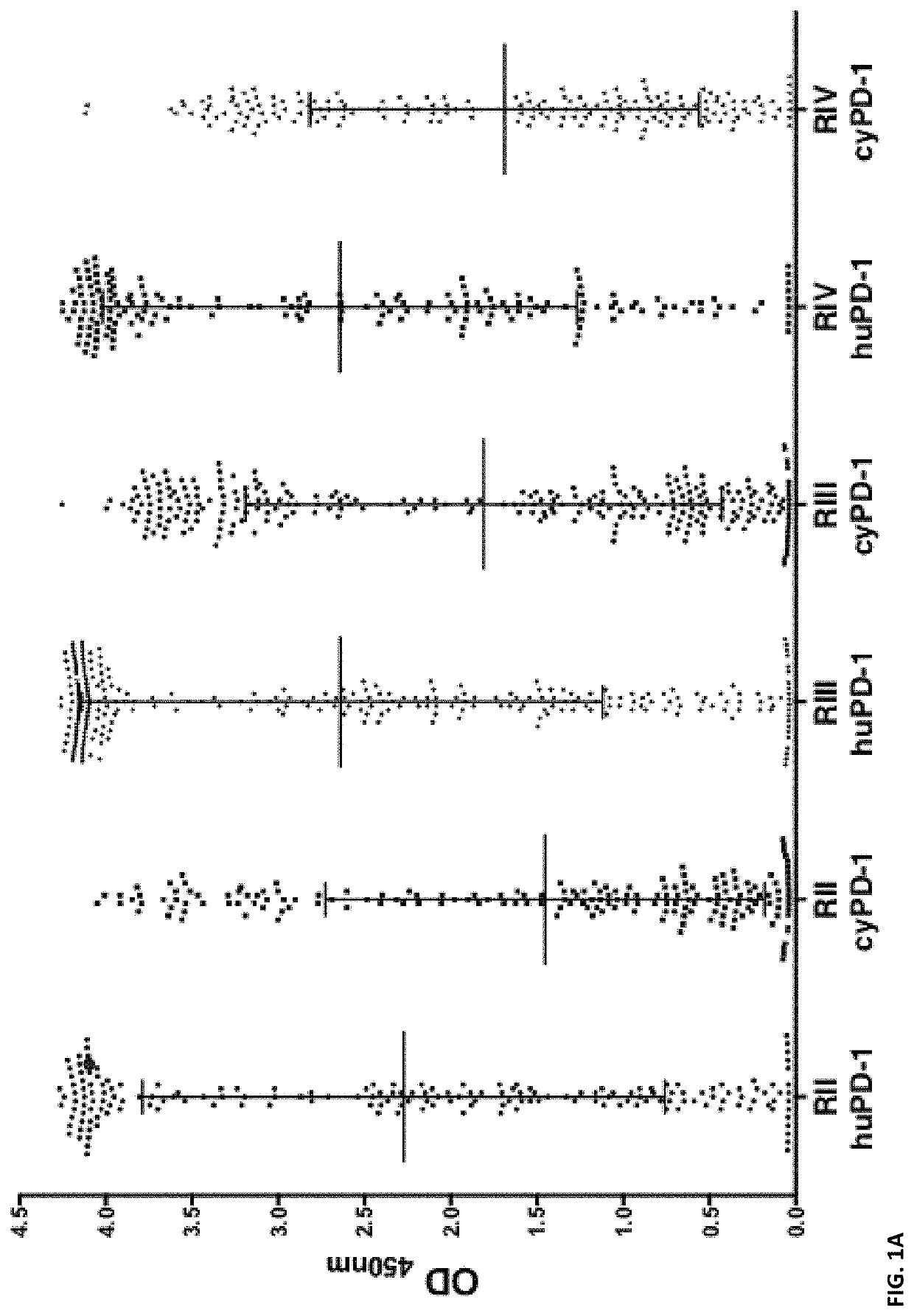
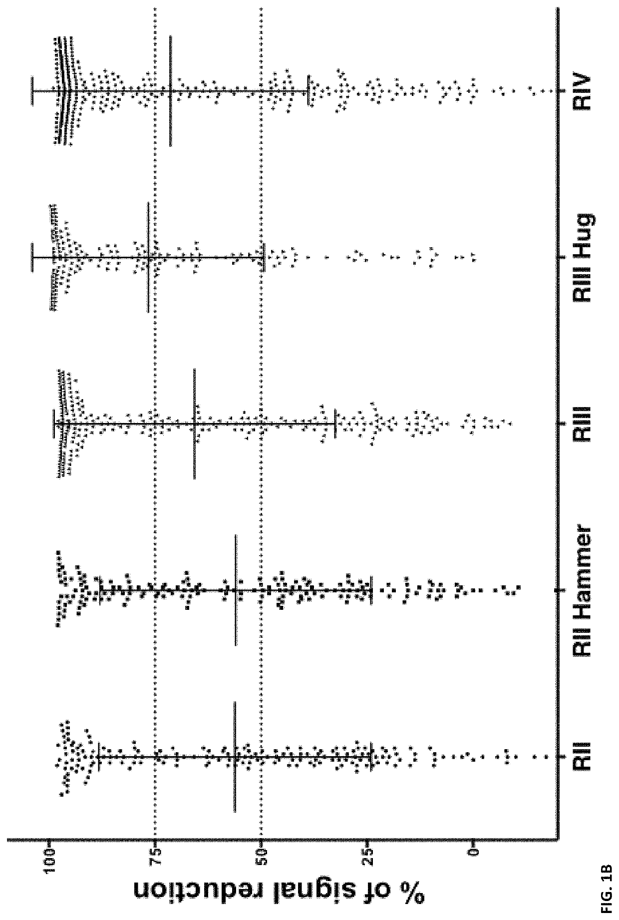
[0297]In this example, we successfully generate a panel of agonistic, optimized anti-PD1 antibodies. These anti-PD1 antibodies are well expressed, biophysically stable, highly soluble and of maximized identity to preferred human germlines.

[0298]Materials and Methods

[0299]PD1 Library Generation and Selection

[0300]The PD1 Fab library was assembled by mass oligo synthesis and PCR. The amplified Fab repertoire was then cloned via restriction-ligation into a phagemid vector, transformed into E. coli TG-1 cells, and the phage repertoire rescued essentially as previously described in detail (Finlay et al., 2011, Methods Mol Biol 681: 383-401).

[0301]Phage selections were performed by coating streptavidin magnetic microbeads with biotinylated PD1 target protein (either human or cyno), washing the beads thrice with PBS and resuspending in PBS pH7.4 plus 5% skim milk protein. These beads were coated at 100 nM target protein in rou...


PUM

PropertyMeasurementUnit
concentrationaaaaaaaaaa
temperatureaaaaaaaaaa
volumeaaaaaaaaaa

Abstract

Provided herein are antibody molecules that bind specifically to Programmed cell death 1 (PD1) and related nucleic acid molecules, vectors and host cells. Also provided herein are medical uses of such antibody molecules.

Description

CROSS-REFERENCE TO RELATED APPLICATIONS[0001]This application is a continuation of International Patent Application No. PCT / EP2019 / 055927, filed on Mar. 8, 2019, which claims the benefit of GB Patent Application No. 1813405.6, filed on Aug. 16, 2018, and GB Patent Application No. 1803746.5, filed on Mar. 8, 2018, the disclosure of each of which is hereby incorporated by reference in its entirety.DESCRIPTION OF THE TEXT FILE SUBMITTED ELECTRONICALLY[0002]The contents of the text file submitted electronically herewith are incorporated herein by reference in their entirety: A computer readable format copy of the Sequence Listing (filename: ULNL_001_02US_SeqList_ST25.txt, date recorded: Jun. 25, 2020, file size ˜161,113 bytes).FIELD OF THE INVENTION[0003]The invention relates to antibody molecules binding specifically to PD1 (also known as Programmed cell death 1, PDCD1, CD279, PD-1, SLEB2, PD-I, SLE1) and medical uses thereof.BACKGROUND OF THE INVENTION[0004]PD1 is a cell surface recep...

Claims


Application Information

Patent Timeline
Patent Type & Authority Patents(United States)
IPC IPC(8): C07K16/28
CPCC07K16/2818C07K2317/76C07K2317/24C07K2317/55C07K2317/33C07K2317/94C07K2317/565C07K2317/52A61P35/00C07K2317/10
Inventor FINLAY, WILLIAM JAMES JONATHAN
Owner ULTRAHUMAN EIGHT LTD
Why Eureka
Try Eureka