Automated methods for the objective quantification of retinal characteristics by retinal region and diagnosis of retinal pathology

a technology of objective quantification and retinal characteristics, applied in the field of clinical diagnosis and monitoring of ocular pathologies utilizing spectral domain optical coherence tomography, can solve the problems of limited objective quantitative data, inability to fully capture, and inability to interpret sd-o

Active Publication Date: 2021-01-12
UNIV OF LOUISVILLE RES FOUND INC
View PDF13 Cites 2 Cited by
  • Summary
  • Abstract
  • Description
  • Claims
  • Application Information

AI Technical Summary

Benefits of technology

[0011]Accordingly, the present disclosure provides a segmentation algorithm that overcomes these and other deficiencies in the art. Methods for quantifying retinal characteristics and diagnosing ocular pathology are also disclosed. Embodiments of the invention are objective and systematic and may be fully automated and executed by a computer, providing reliable and robust diagnoses and monitoring of even occult retinal conditions and pathologies.

Problems solved by technology

However, with the exception of retinal thickness measurements, conventional methods relying on visual inspection of SD-OCT provides limited objective quantitative data, and therefore images must be subjectively interpreted by an eye specialist.
As a result, interpretation of SD-OCT is susceptible to human bias and error.
The current techniques for observing and analyzing the aging retina in vivo, including longitudinal OCT imaging in high-risk populations, fail to fully capture the subtle changes that occur such that even with monitoring, disease is not typically diagnosed until irreversible damage has occurred.
Although initial preliminary automated image processing exists in some commercially available SD-OCT models, it is currently limited to retinal thickness, retinal volume and partial retinal segmentation.
This algorithm failed with poor-quality images and also failed with some good-quality ones.
However, further work is needed to apply this algorithm to more advanced retinal abnormalities.
The algorithm, however, could not separate highly reflective image features.
This method performed well even when retinal blood vessels shaded the layers, but failed in blurry images.
This algorithm showed promise in segmenting both healthy and diseased scans; however has not demonstrated reliable evaluation on retinas affected with outer / inner retinal diseases.
However, the algorithm worked only with high-contrast images.
The above discussion demonstrates that there are limitations associated with known retinal layer segmentation OCT-based protocols, for example, low accuracy achieved when having images with low signal to noise ratio (SNR), and the fact that the majority of the proposed approaches could segment only up to eight retinal layers, while methods that segmented more layers were successful only with high-contrast images.

Method used


Image

  • Automated methods for the objective quantification of retinal characteristics by retinal region and diagnosis of retinal pathology
  • Automated methods for the objective quantification of retinal characteristics by retinal region and diagnosis of retinal pathology
  • Automated methods for the objective quantification of retinal characteristics by retinal region and diagnosis of retinal pathology

Examples


example 1

[0062]This example illustrates development of a retinal SD-OCT segmentation algorithm in accordance with embodiments of the invention.

A. Data Collection

[0063]Subjects were recruited at the Kentucky Lions Eye Center, University of Louisville Department of Ophthalmology and Visual Sciences, Louisville, Ky. between June 2015 and December 2015. Informed consent was obtained from all participants. Subjects with normal retinas ranging in age from 10 to 79 years old were included in the study. Past medical history, ophthalmologic history, smoking status, and current medications were obtained via chart review and subject interview. Persons with history significant for any retinal pathology, history significant for diabetes mellitus, high myopia defined as a refractive error greater than or equal to 6.00 diopters, and tilted OCT image were excluded from participation in the study. SD-OCT scans were prospectively collected from 165 noiuial subjects using the Zeiss Cirrus HD-OCT 5000. SD-OCT d...

example 2

[0094]This example illustrates derivation of quantitative data from segmented SD-OCT images in accordance with an embodiment of the invention. In particular the concept is illustrated utilizing reflectivity as the quantified characteristic and development of a normalized reflectivity scale.

Quantitative Data Derived from SD-OCT Images

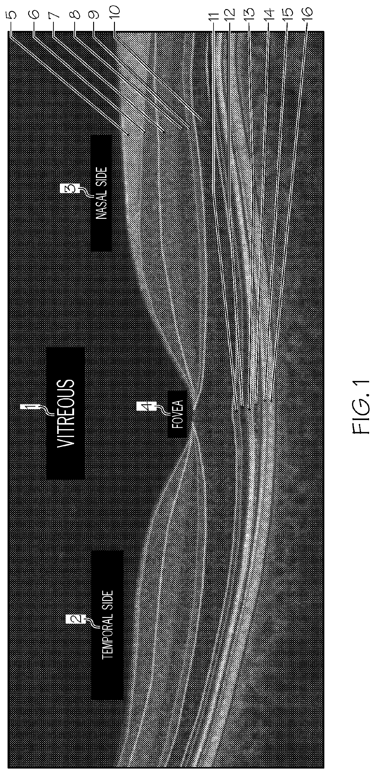
[0095]Several quantitative data can be derived from the segmented SD-OCT images in order to optimally characterize retinal morphology. Specific embodiments herein are described in terms of the reflectivity measurements that were obtained from two regions per scan, comprising the thickest portions of the retina on the nasal and temporal sides of the foveal peak. Although the embodiment is described and illustrated utilizing reflectivity, it will be apparent to the person of skill in the art that the methods are applicable for other quantifiable retinal characteristics.

[0096]Mean reflectivity is expressed on a normalized scale, calibrated such that the for...

example 3

[0097]This example illustrates application of an embodiment of the automated segmentation algorithm to an analysis of retinal layer reflectivity as a function of gender, age, and demographic. The reflectivity unit (RU) data is summarized by age in FIG. 9.

[0098]After the segmentation protocol is applied to the collected images, analysis of the reflectivity measure across the different decades of life is undertaken. Embodiments of the segmentation approach were first validated using “ground truth” for subjects, which was collected from 165 subjects aged 10-79 years. Subjects with high myopia (≤−6.0 diopters), and tilted OCT were excluded. This ground truth was created by manual delineations of retina layers reviewed with 4 different retina specialists (SS, AP, AH, DS).

[0099]This study was reviewed and approved by the Institutional Review Board at the University of Louisville. A retrospective chart review was performed to identify subjects between 13 and 97 years of age without signifi...


PUM


Abstract

Automated and objective methods for quantifying a retinal characteristic include segmenting an optical coherence tomography image into a plurality of layered retinal regions, and quantifying the retinal characteristic for each region as normalized to a range defined by the characteristic value in the vitreous region and in the retinal pigment epithelium region. Such methods are useful for detecting occult ocular pathology, diagnosing ocular pathology, reducing age-bias in OCT image analysis, and monitoring efficacy ocular / retinal disease therapies.

Description

CLAIM OF PRIORITY[0001]This application claims priority to U.S. Provisional Applicant No. 62 / 256,980 filed on Nov. 18, 2015, the entire disclosure of which is incorporated herein by this reference.TECHNICAL FIELD[0002]The present disclosure relates to the field of clinical diagnosis and monitoring of ocular pathologies utilizing spectral domain optical coherence tomography (SD-OCT). Specifically, the disclosure relates to automated methods for segmenting and analyzing an OCT image and automated methods for diagnosing ocular pathology based thereon.BACKGROUND ART[0003]Spectral domain optical coherence tomography (SD-OCT) is a widely used tool for the diagnosis and evaluation of retinal diseases. Utilizing interferometry, low coherence light is reflected from retinal tissue to produce a two-dimensional grayscale image of the retinal layers. Differences in reflectivity of retinal layers produce different intensities on an SD-OCT scan, allowing for noninvasive visualization of distinct ...

Claims


Application Information

Patent Timeline
Patent Type & Authority Patents(United States)
IPC IPC(8): G06K9/00G06T7/00G06T7/12G06T7/143G06T7/168A61B3/10G06T7/149
CPCG06T7/0012A61B3/102G06T7/12G06T7/143G06T7/149G06T7/168G06T2207/10101G06T2207/20064G06T2207/20121G06T2207/20124G06T2207/30041
Inventor SCHAAL, SHLOMITEL-BAZ, AYMANHARJRASOULIHA, AMIR REZA
Owner UNIV OF LOUISVILLE RES FOUND INC
Why Eureka
Try Eureka