Apparatus and method for enhanced early photon detection in optical projection tomography

an optical projection tomography and early photon detection technology, applied in the field of medical imaging using optical projection tomography, can solve the problem of high scattering nature of optical projection tomography photon propagation in biological tissue, and achieve the effect of improving spatial resolution

Active Publication Date: 2021-04-27
ILLINOIS INSTITUTE OF TECHNOLOGY
View PDF8 Cites 1 Cited by
  • Summary
  • Abstract
  • Description
  • Claims
  • Application Information

AI Technical Summary

Benefits of technology

[0004]The invention improves existing “early photon” optical projection tomography (both absorption and fluorescence based), through use of innovativ...

Problems solved by technology

However, a major limitation to optical tomography is the hi...

Method used


Image

  • Apparatus and method for enhanced early photon detection in optical projection tomography
  • Apparatus and method for enhanced early photon detection in optical projection tomography
  • Apparatus and method for enhanced early photon detection in optical projection tomography

Examples


examples

[0040]The present invention is described in further detail in connection with the following examples which illustrate or simulate various aspects involved in the practice of the invention. It is to be understood that all changes that come within the spirit of the invention are desired to be protected and thus the invention is not to be construed as limited by these examples.

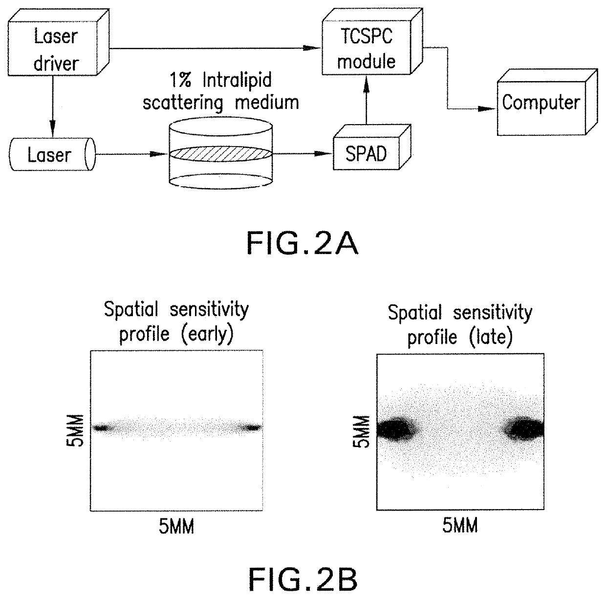
[0041]FIG. 2A illustrates an experimental setup of an apparatus according to one embodiment of this invention. FIG. 2B shows the simulated spatial sensitivity profile as observed with only early arriving photons or the ballistic and quasi ballistic photons vs. the late arriving ones which are diffused in the medium and have suffered scattering losses. The experimental setup was built around a single excitation laser at 785 nm (LDH-PC780 and PDL 800-B laser driver, Picoquant, USA) and one state-of-the-art time-correlated single photon counting (TCSPC) (Picoharp 300, Picoquant, USA) single photon avalanche diode (S...


PUM


Abstract

A system and method for optical tomography including illuminating an object with pulsing stimulus light and pulsing the stimulus light at a repetition frequency having a pulse period that is greater than a dead-time of a detector. Coordinating the pulse with the dead-time of the detector allows for higher powered light source and improves early photon detection.

Description

CROSS REFERENCE TO RELATED APPLICATION[0001]This application claims the benefit of U.S. Provisional Patent Application Ser. No. 62 / 327,800, filed on 26 Apr. 2016. The Provisional Application is hereby incorporated by reference herein in its entirety and is made a part hereof, including but not limited to those portions which specifically appear hereinafter.BACKGROUND OF THE INVENTION[0002]This invention relates generally to medical imaging using optical projection tomography and, more particularly, to a method and apparatus capable of achieving high-resolution, quantitative mapping of fluorescence targeted biological molecular concentration distributions, such as in 1-10 mm diameter tissues.[0003]Absorption and fluorescence-based optical tomography has been heralded as a low-cost, ionizing radiation-free alternative to conventional medical imaging modalities for decades, particularly for tissue specimen and small animal imaging. However, a major limitation to optical tomography is t...

Claims


Application Information

Patent Timeline
IPC IPC(8): A61B5/00G01N21/64H01L31/107
CPCA61B5/0073A61B5/0071G01N21/64H01L31/107A61B2562/0233A61B2576/00G01N21/6456G01N2201/0697G01N2201/06113G01N2201/12G16H30/40
Inventor TICHAUER, KENNETH M.BRANKOV, JOVAN G.SINHA, LAGNOJITA
Owner ILLINOIS INSTITUTE OF TECHNOLOGY
Why Eureka
Try Eureka