Instrument for accessing and visualizing hollow organs

a hollow organ and instrument technology, applied in the field of hollow organ accessing and visualizing instruments, can solve the problems of unintentional tightening of teeth by patients during endoscopic examination, affecting the patient's experience of laryngobronchoscopy, so as to avoid the onset of coughing and choking sensation, minimize discomfort and pain, and reduce discomfort and pain

Active Publication Date: 2022-01-18
FOND PER LA CURA MINI INVASIVA TUMORI ONLUS
View PDF16 Cites 0 Cited by
  • Summary
  • Abstract
  • Description
  • Claims
  • Application Information

AI Technical Summary

Benefits of technology

[0019]The flexible tubular element it is provided with a locking system at its proximal end which allows to attach it to the mouthpiece when its distal end has been correctly positioned in the esophagus or trachea. According to a preferred embodiment of the invention, the flexible tubular element is removably inserted into the sleeve of the mouthpiece, thus allowing to simplify the structure of the individual components and to use interchangeable flexible tubular elements of different lengths according to age, sex, the size of the oral cavity and the anatomical conformation of the patient.
[0030]In addition to the aforementioned advantages, with respect to the use of flexible laryngoscopes as mandrels for guiding an anesthesia tube, the instrument according to the invention has the advantage of not requiring the use of endoscopic columns in the operating room, especially in emergency procedures performed outside an operating room. Furthermore, it is not necessary to sterilize the endoscope as it is supplied sterile. Another advantage is that the flexible tubular element bound to the mouthpiece allows continuous vision of the trachea and lobar bronchi during an operation and the possible targeted aspiration of the bronchial secretions. A further advantage offered by the invention is that the instrument for accessing and visualizing hollow organs is in principle structurally simpler and less expensive than the endoscopy instruments currently available. Other advantages, features and methods of use of the present invention will become evident from the following detailed description of some embodiments thereof, presented for illustrative and non-limiting purposes.

Problems solved by technology

As known, the introduction of an endoscope into a patient's oral cavity exposes the endoscope itself to the risk of bite damage.
In fact, the patient during the endoscopic examination can unintentionally tighten the teeth and damage the sheath of the instrument.
Despite these measures, the esophagus-gastro-duodenoscopy and laryngo-bronchoscopy are invasive examinations which are rather annoying for a patient, when introducing the endoscope into the oral cavity, when advancing the instrument in the hollow organs to be inspected and when sampling tissue samples.
On the other hand there is the profuse salivation that induces in the patient the involuntary reflex of swallowing, which is however hindered and made impossible by the presence of the endoscope.
This results in the feeling of nausea and retching and the unwanted ab-ingestis which induces persistent cough and a sense of suffocation.
The common problems related to the use of rigid laryngoscopes are related to the traumatism due to their introduction into the oral cavity, which, having to be rapid due to the apnea induced by the drugs administered in the induction phase of the anesthesia, determines trauma and damage of the lips, tongue, teeth, gums and vocal cords which result in permanent damage causing civil or criminal cases with a claim for compensations.
The risk of non-intubation in the induction phase of anesthesia can cause severe and persistent hypoxia with possible neurological and even fatal damage.
In cases of difficult intubation, the use of the flexible laryngoscope requires a programmed displacement to an operating room of an endoscopic column, which is expensive and not always available, and the use of a non-dedicated optical vision system that requires sterilization.

Method used


Image

  • Instrument for accessing and visualizing hollow organs
  • Instrument for accessing and visualizing hollow organs
  • Instrument for accessing and visualizing hollow organs

Examples


Embodiment Construction

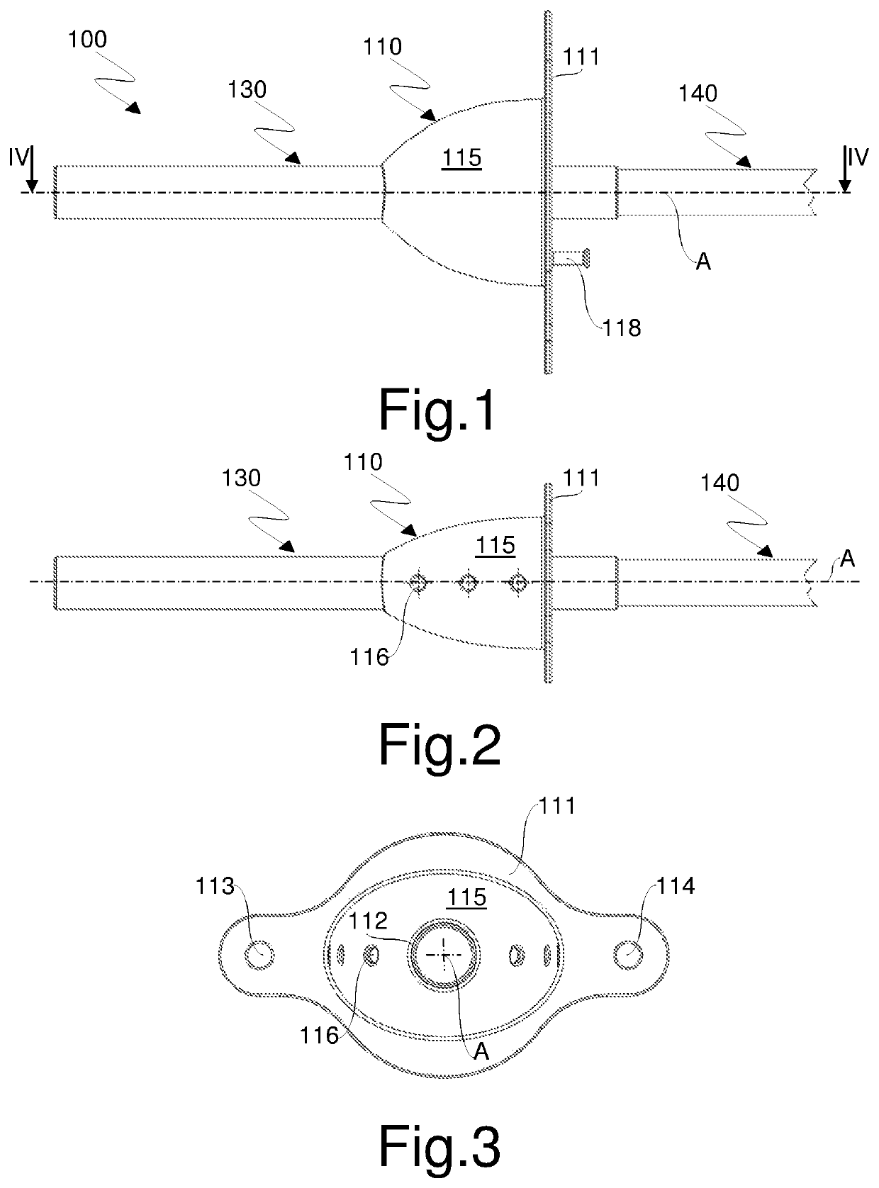
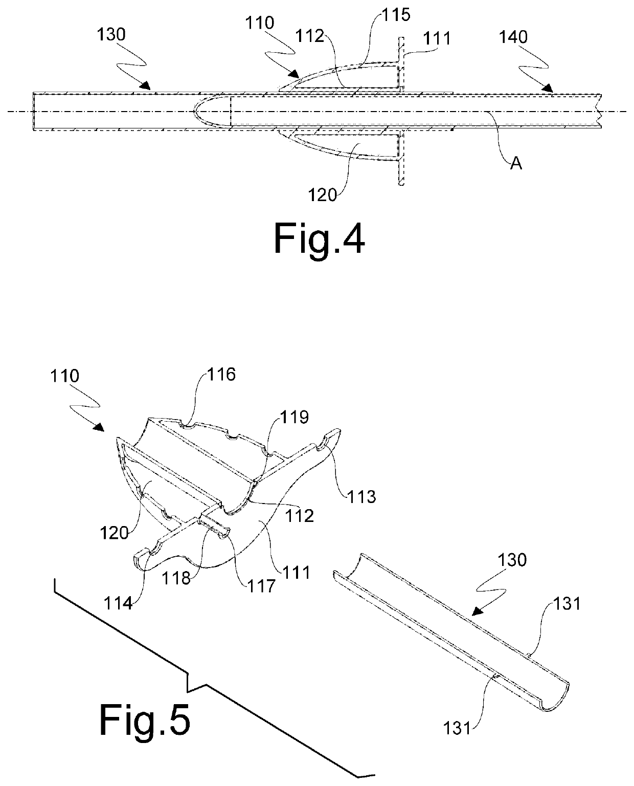
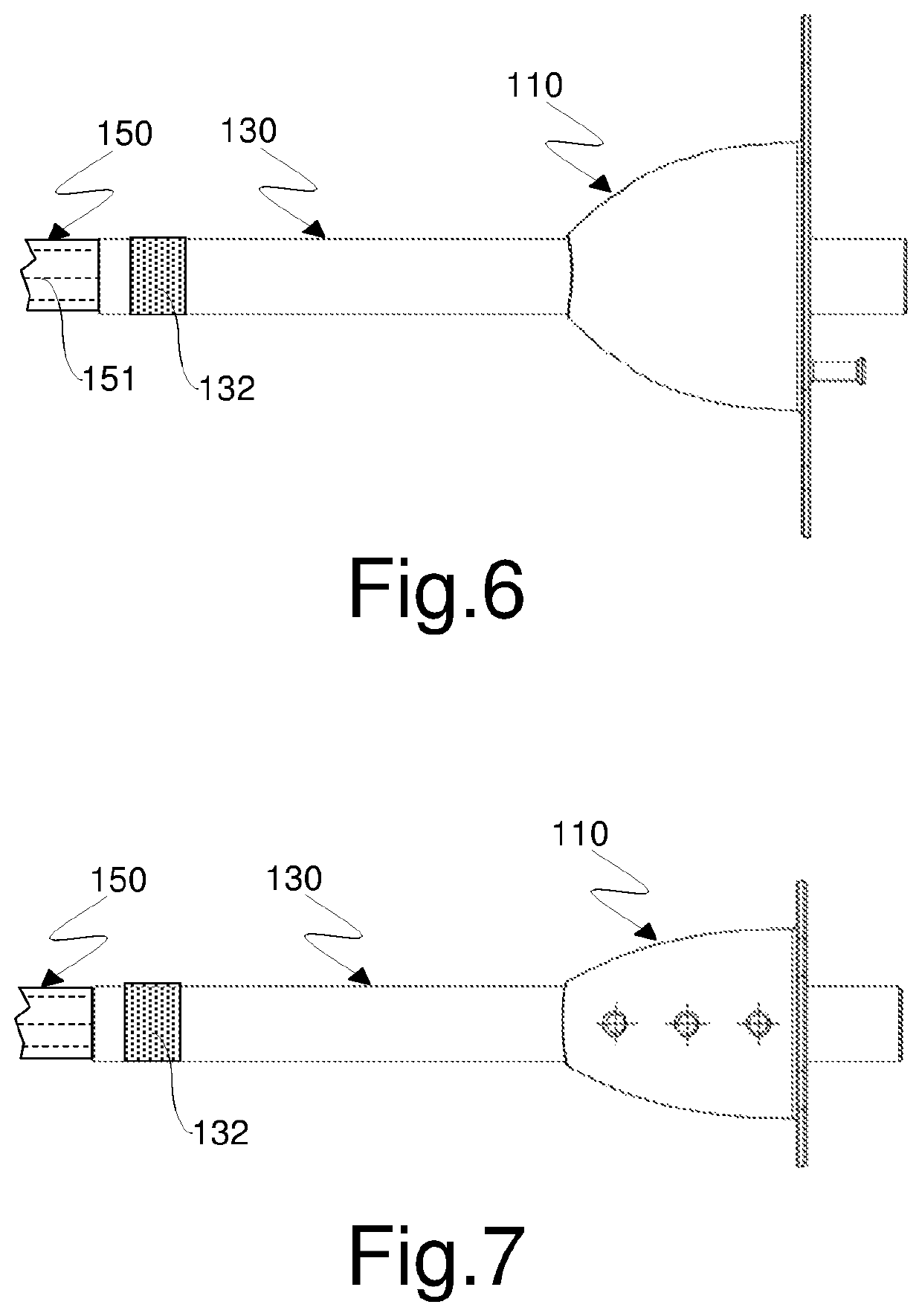
[0038]With reference initially to FIGS. 1 to 5, an instrument for accessing and visualizing hollow organs according to the invention is generally indicated by reference numeral 100.

[0039]Instrument 100 comprises a mouthpiece 110 configured to be inserted into a patient's mouth. The mouthpiece 110 comprises, in a known manner, a mask 111 which is perforated, for example in the center, and a sleeve 112 having a generally cylindrical shape and a circular or elliptical cross-section which extends from the hole formed in the mask 111 in a direction substantially perpendicular to it, thereby defining a channel suitable for allowing the introduction of a flexible tubular element. The mask 111 also comprises in a known manner a pair of through openings 113, 114, for example of circular shape, obtained at its opposite ends with respect to an axis A of the sleeve 112 and configured to allow the assembly of a band (not shown) which is elastic or of another suitable material for maintaining the...


PUM


Abstract

An instrument for accessing and visualizing hollow organs is provided. The instrument includes a mouthpiece having a perforated mask and a sleeve having a generally cylindrical or ovoid shape extending from the perforated mask substantially perpendicularly thereto and defining a channel. The instrument further includes sucking means having a suction chamber or closed volume that is restrained to the mask on the side where the sleeve is formed. The suction chamber has a plurality of through openings adapted to allow fluid communication with a surrounding environment, an intake opening communicating with the suction chamber formed on the mask, wherein the through openings are formed on the skirt of a dome-shaped member which delimits the suction chamber.

Description

CROSS-REFERENCE TO RELATED APPLICATIONS[0001]This application is a 371 of PCT / IB2018 / 052775, filed Apr. 20, 2018, which claims the benefit of Italian Patent Application No. 102017000046337, filed Apr. 28, 2017.TECHNICAL FIELD OF THE INVENTION[0002]The present invention relates in general to devices for accessing and visualizing hollow organs and in particular an instrument which can be used for guiding endoscopes in esophagus-gastro-duodenoscopy and laryngo-bronchoscopy procedures. The instrument according to the invention can also be used for the intubation and the ventilation of a patient in general anesthesia procedures performed both in the election and in the emergency regimes.BACKGROUND[0003]The esophagus-gastro-duodenoscopy is an instrumental examination performed by specialized doctors to diagnose, and in selected cases treat, pathologies of the upper gastro-enteric tube. The examination is performed by introducing an endoscope into a patient's oral cavity and advancing its ...

Claims


Application Information

Patent Timeline
Patent Type & Authority Patents(United States)
IPC IPC(8): A61M16/04A61M16/06
CPCA61M16/0495A61M16/0463A61M16/0465A61M16/0477A61M16/0493A61M16/0627A61M2202/0466A61M2205/583A61B1/00154A61B1/015A61B1/273A61B90/16A61B1/2676A61B1/01A61M16/0488A61M16/0486
Inventor GARBAGNATI, FRANCESCO ANTONIOMARIA
Owner FOND PER LA CURA MINI INVASIVA TUMORI ONLUS
Why Eureka
Try Eureka