Compositions and methods of cell attachment

a cell attachment and cell technology, applied in the field of cell attachment, can solve the problems of often applying generically with mixed ecms

Active Publication Date: 2022-11-22
EMULATE INC
View PDF21 Cites 0 Cited by
  • Summary
  • Abstract
  • Description
  • Claims
  • Application Information

AI Technical Summary

Benefits of technology

[0036]The term “microfluidic” as used herein relates to components where moving fluid is constrained in or directed through one or more channels wherein one or more dimensions are 1 mm or smaller (microscale). Microfluidic channels may be larger than microscale in one or more directions, though the channels) will be on the microscale in at least one direction. In some instances the geometry of a microfluidic channel may be configured to control the fluid flow rate through the channel (e.g. increase channel height to reduce shear). Microfluidic channels can be formed of various geometries to facilitate a wide range of flow rates through the channels.

Problems solved by technology

However, these ECMs are often applied generically with mixed results, depending on the cell type and culture conditions.

Method used


Image

  • Compositions and methods of cell attachment
  • Compositions and methods of cell attachment
  • Compositions and methods of cell attachment

Examples


example 1

Cellular Crosslinking to Improve Cell Attachment to Channels

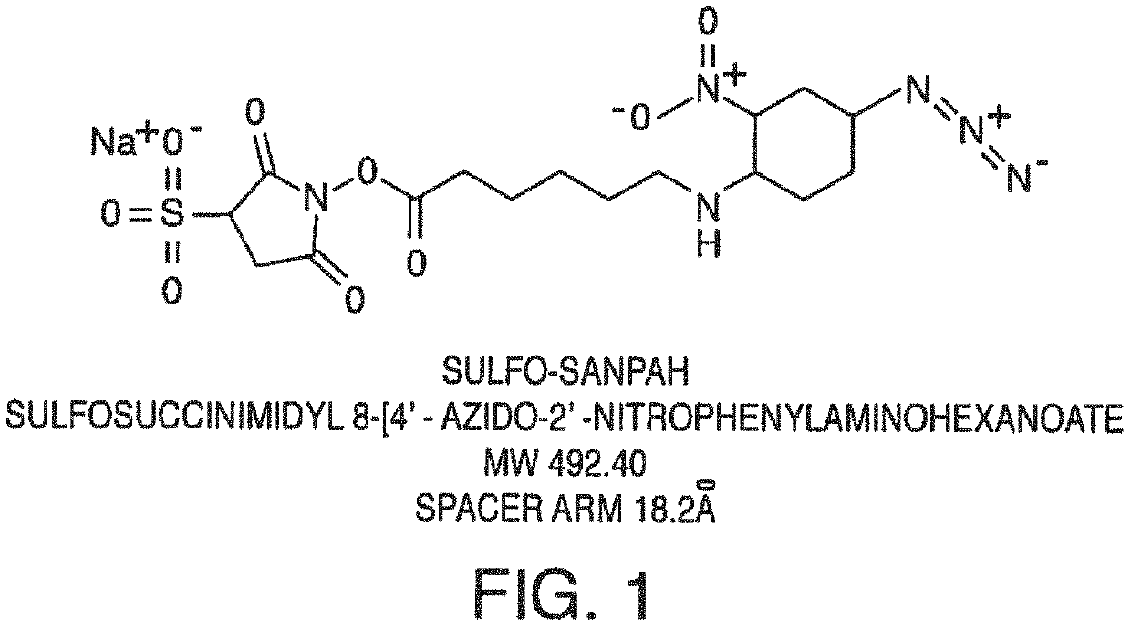
[0119]In one embodiment, the present invention contemplates using a crosslinker to covalently attach proteins or peptides that enhance cell attachment. In this example, a protocol for using Sulfa-SANPAH as the crosslinker is provided as one embodiment of a method.

[0120]First, fresh 0.5 mM Sulfo-SANPAH (492.4 g / mol) solution in 50 mM HEPES (0.22 um sterile filtered, pH 7.4) (protect from light) is prepared. Then, an ECM solution is prepared (e.g. 50 ug / mL Laminin in PBS or media without FBS) on ice.

[0121]The microfluidic device (“chip”) comprising a microchannel is then plasma treated. Plasma—15 sccm O2, 60 sec, 100 W.

[0122]The channels are then washed with 200 uL of 50 mM HEPES. Excess 50 mM HEPES is removed from the channel.

[0123]Sulfo-SANPAH is introduced into the microchannel by inserting a pipet tip reservoir in a port of the chip. 100 uL of Sulfo-SANPAH solution is added to the top channel, ejecting tip into inlet port...

example 2

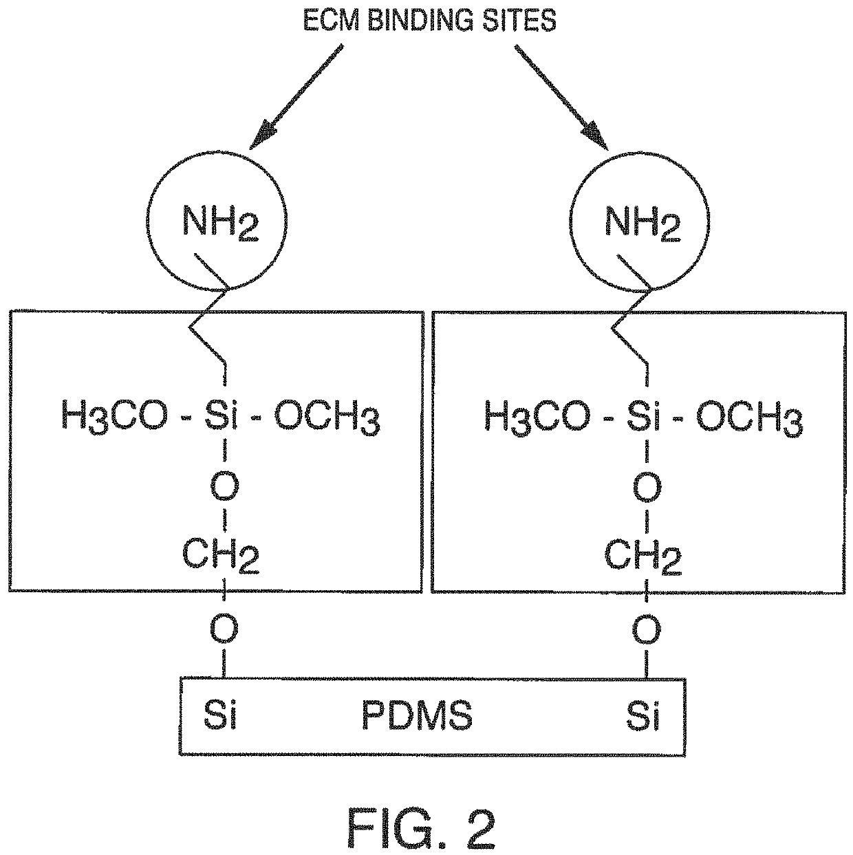
Channel Surface Modification to Improve Cell Attachment

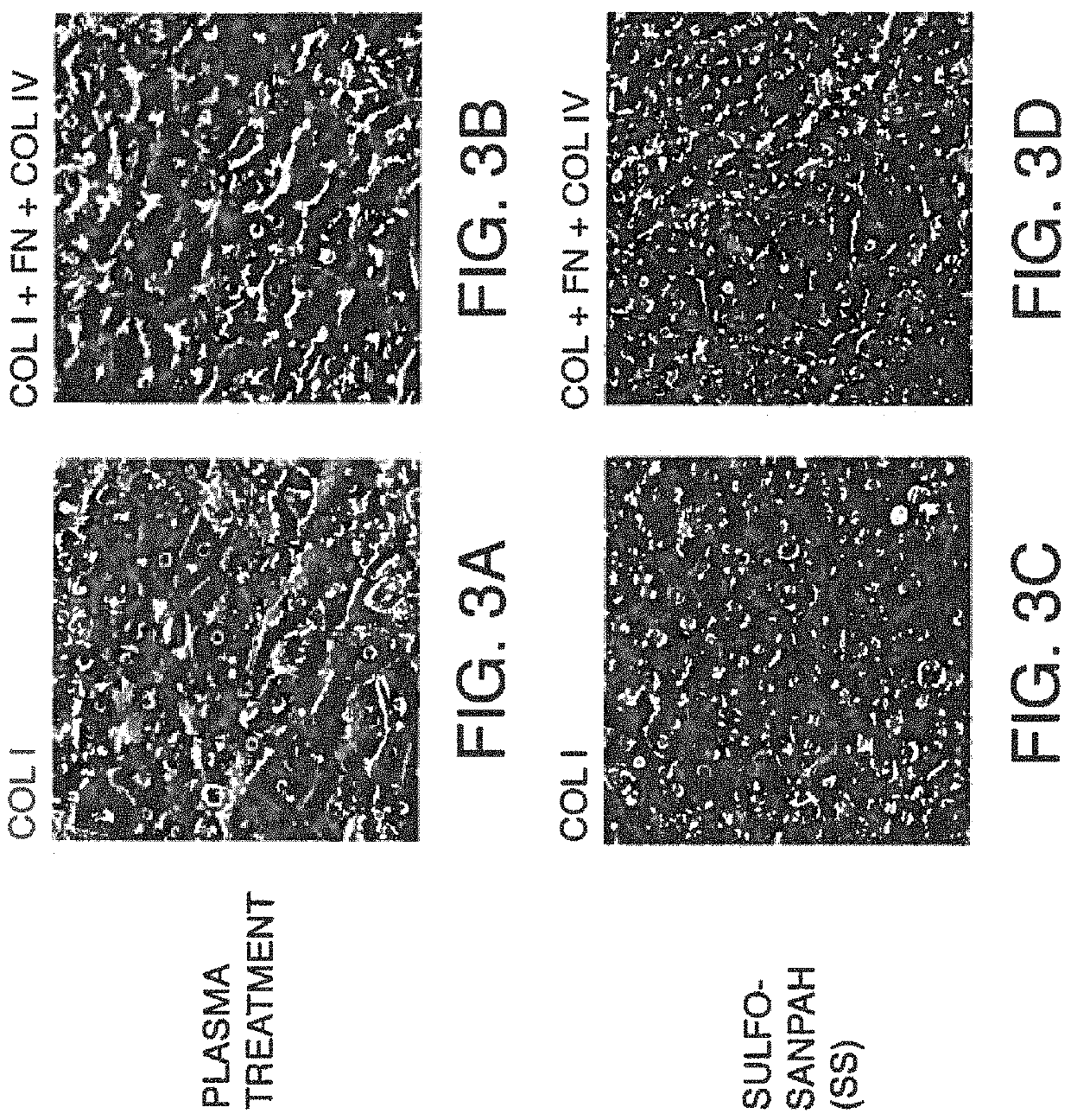
[0127]In this example, PDMS surfaces treated with plasma were compared with PDMS surfaces modified by covalent attachment ECM proteins. FIGS. 3A-B show photographs of hepatocytes six (6) days after being seeded on a PDMS surface that was either plasma treated (FIGS. 3 A & B) or that was Sulfo-SANPAH treated (i.e. ECM protein(s) covalently attached to the surface with this crosslinker) (FIGS. 3 C & D). The cells were cultured under flow conditions for two (2) days.

[0128]FIGS. 4A-D show photographs of hepatocytes nine (9) days after being seeded on a PDMS surface that was either plasma treated (FIGS. 4A& B) or that was Sulfo-SANPAH treated (i.e. ECM protein(s) covalently attached to the surface with this crosslinker) (FIGS. 4C& D). The cells were cultured under flow conditions for 5 days.

[0129]FIGS. 5A-B show photographs of hepatocytes fourteen (14) days after being seeded on a PDMS surface that was either plasma treated (FIG. 5A)...

example 3

Shelf-Life Study of ECM-Coated Chips

[0130]This example evaluates conditions to avoid ECM (i.e., for example, laminin, Matrigel) inactivation found during dry storage. All tested chips were stored at 4° C. then compared to freshly coated chips. Results indicate that ECM for Gut-on-Chip chips are best stored in solution (e.g., wet).

Experimental Design

[0131]The ECM for Gut-on Chip chips comprised Matrigel and collagen I. Gut-on-Chip chips were chosen as test platform due to its robustness to varying culture conditions. In particular, test Chips were treated with Sulfo-SANPAH and ECM (100 ug / mL Martigel and 25 ug / mL collagen I). All conditions were compared to freshly coated chips.

Results

[0132]Twenty-eight (28 chips) were stored for 1 week. No differences in Caco-2 and HUVEC cell attachment was observed. No differences in Caco-2 and HUVEC cell morphology was observed. The chips were maintained for 8 days prior to exposure to TNF-α and IL-1β. The experiment also included some chips with ...


PUM

PropertyMeasurementUnit
temperatureaaaaaaaaaa
wavelengthsaaaaaaaaaa

Abstract

Compositions, devices and methods are described for improving adhesion, attachment, and / or differentiation of cells in a microfluidic device or chip. In one embodiment, one or more ECM proteins are covalently coupled to the surface of a microchannel of a microfluidic device. The microfluidic devices can be stored or used immediately for culture and / or support of living cells such as mammalian cells, and / or for simulating a function of a tissue, e.g., a liver tissue, muscle tissue, etc. Extended adhesion and viability with sustained function over time is observed.

Description

FIELD OF THE INVENTION[0001]The present invention contemplates compositions, devices and methods of improving adhesion, attachment, and / or differentiation of cells in a microfluidic device or chip. In one embodiment, ECM protein(s) is covalently coupled to the surface of a microchannel of a microfluidic device. The microfluidic devices can be either stored, or immediately used for culture and / or support of living cells such as mammalian cells, and / or for simulating a function of a tissue, e.g., a liver tissue, muscle tissue, etc. Extended adhesion and viability with sustained function over time is observed.BACKGROUND[0002]Cell adhesion is a central mechanism that ensures the structural integrity of tissue and is often a requirement for its biological function. The most prominent cell-matrix adhesion structures are so-called focal contacts. Focal contacts consist of large patches of transmembrane adhesion receptors from the integrin-family. These integrin patches in the cell membrane...

Claims


Application Information

Patent Timeline
Patent Type & Authority Patents(United States)
IPC IPC(8): C12N5/00C12N5/02A61K38/00C07K2/00C07K4/00C07K5/00C07K7/00C07K14/00C07K16/00C07K17/00G01N33/50C07K14/78C12N5/071C12M3/06C12M1/00C12M1/12C12N5/077
CPCG01N33/5032C07K2/00C07K14/78C12M23/16C12M23/20C12M25/02C12N5/0018C12N5/0068C12N5/067C12N5/0658G01N33/5014C12N2500/32C12N2521/00C12N2533/30C12N2533/50C12N2533/52C12N2533/54C12N2533/90C12N2535/10C12N2537/10G01N33/5044
Inventor LEVNER, DANIELJANG, KYUNG JINFRASER, JACOBKERNS, S. JORDANVARONE, ANTONIOHUH, DONGEUN
Owner EMULATE INC
Why Eureka
Try Eureka