
Acousto-thermal shift assay for label-free protein analysis

Pending Publication Date: 2022-09-15
THE REGENTS OF THE UNIV OF COLORADO - A BODY CORP
View PDF0 Cites 0 Cited by
  • Summary
  • Abstract
  • Description
  • Claims
  • Application Information

AI Technical Summary

Benefits of technology

This patent is about a technology called microfluidic that involves using small channels to control the movement of fluids. These channels can be very small and can have specific designs to help control how fast the fluid moves through them. This technology can be used to create components that can manage small amounts of fluid in a precise way.

Problems solved by technology

However, conventional measurement assays and sample preparation takes from many hours up to a whole day and / or requires bulky / expensive facility and experiences.
This limitation is one of the main challenges that lead to high cost and long development cycles for therapeutics and drug development in pharmaceutical industry and particularly in the development of portable medical devices for fast and low cost diagnosis and healthcare.

Method used


Image

  • Acousto-thermal shift assay for label-free protein analysis
  • Acousto-thermal shift assay for label-free protein analysis
  • Acousto-thermal shift assay for label-free protein analysis

Examples


example i

Chemicals

[0103]Two proteins, i.e. human hemoglobin (Hb; Millipore Sigma, St. Louis, Mo., USA) and citrate synthase (CS) from porcine heart (Millipore Sigma), and two corresponding compounds, i.e. palmatine chloride (Pal; Santa Cruz Biotechnology, Santa Cruz, Calif., USA) and oxaloacetic acid (OAA; Millipore Sigma) were primarily used. Human whole blood samples with K2EDTA as an anticoagulant were purchased from Zen-Bio Inc. (Research Triangle Park, N.C., USA) and stored in 4° C. (always used within 3-14 days after collection). Human blood plasma was obtained by centrifuging the human whole blood samples at 500 g for 10 min in 4° C. Sickle cell diseased (SCD) red blood cell lysate were obtained from patients with SCD, upon receiving written informed consent and in conformity with the declaration of Helsinki under a protocol approved by the Duke University Medical Center (no. NCT02731157) as described previously. Culp-Hill et al., “Effects of red blood cell (RBC) transfusion on sickle...

example ii

A-TSA Device Fabrication

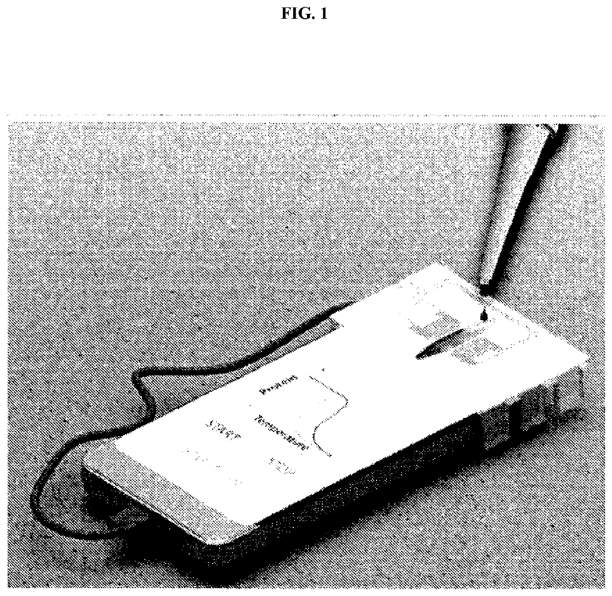
[0104]The SAW was generated and propagated on piezoelectric 128° Y-cut X-propagating lithium niobate (LiNbO3) wafer (500 μm thick). The device consisted of a pair of interdigitated transducers (IDTs) in parallel in order to generate two series of identical SAWs propagating in the opposite direction, producing a standing SAW. Each IDT consists of 30 pairs of electrodes (Cr / Au, 5 / 100 nm) with the width of electrode finger of 50 μm, pitch of 100 μm, and an aperture of 10 mm, yielding a frequency of approximately 20 MHz for the propagating SAW. Although different IDTs can be used, the resonance frequencies of most IDTs are in the range between 19.5 and 19.6 MHz. A PDMS microchannel with height of 100 μm and width of 2 mm was then fabricated through a standard soft-lithography and model-replica procedure. Lastly, both the PDMS channel and the IDT substrate were treated with oxygen plasma and bonded together to form the final SAW device. See, FIG. 4B.

example iii

Acousto-Thermal Shift Assay (A-TSA) and Data Analysis

[0105]The A-TSA SAW device was mounted on the stage of an inverted microscope (ECLIPSE Ti-U, Nikon, Japan). A radio frequency (RF) signal was generated by a function generator (EXG Analog Signal Generator, Keysight, Santa Rosa, Calif., USA) and amplified by an amplifier (403LA, Electronics & Innovation, Rochester, N.Y., USA). Five microliters of protein, plasma, red blood cell lysate or protein-compound mix solutions were injected into the channel before the RF signals were applied. A fast camera (ORCA-Flash4.0LT, Hamamatsu, Japan) was connected to the microscope to capture the process, and all the videos were recorded in 4 frames per second.

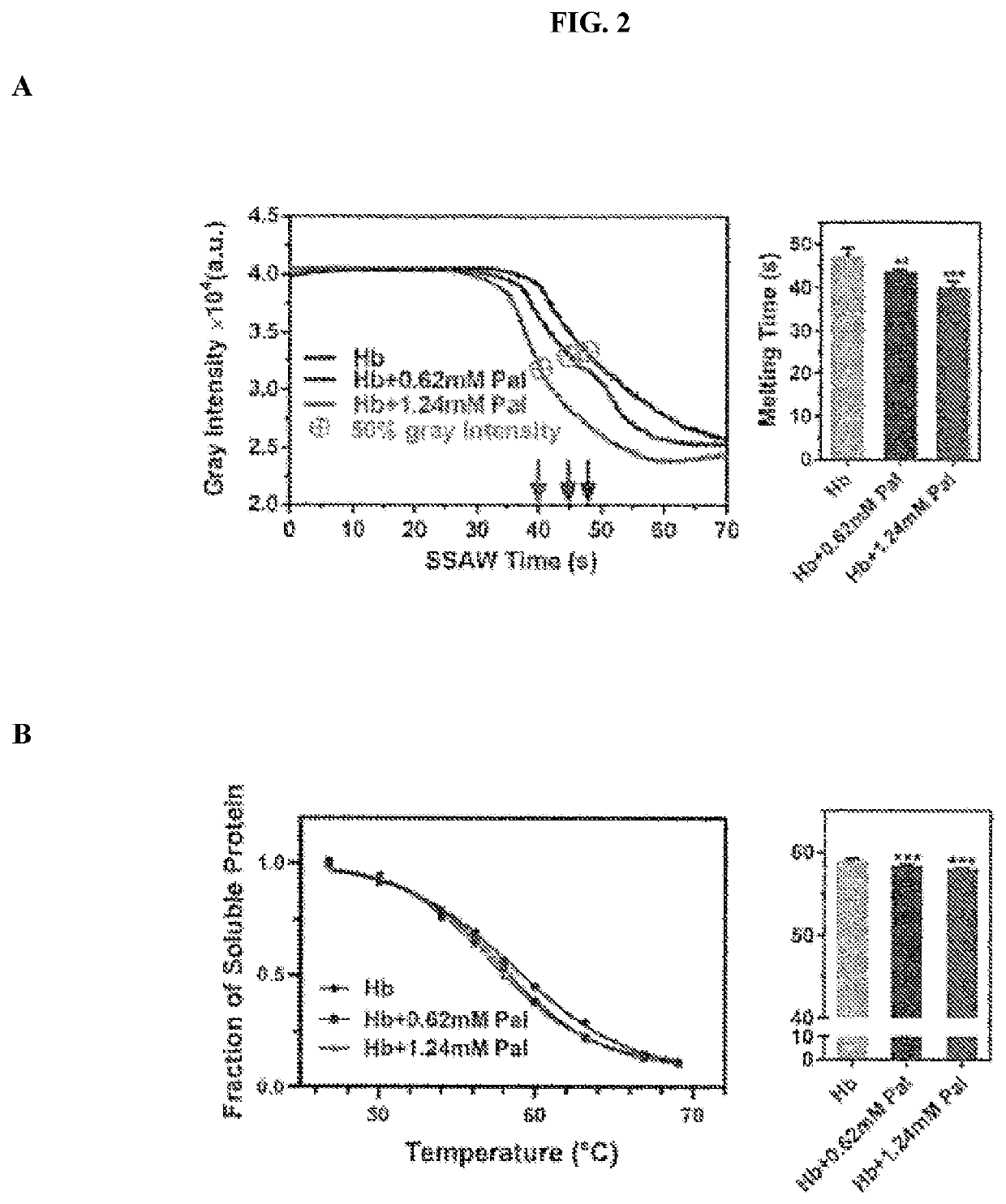
[0106]All image and videos processing were performed in ImageJ (National Institute of Health, Bethesda, Md., USA) in the same way as described below. The same sized regions of interest (ROIs) were traced around the perimeter of each pattered protein fiber in order to monitor the gray intensity...


PUM


Abstract

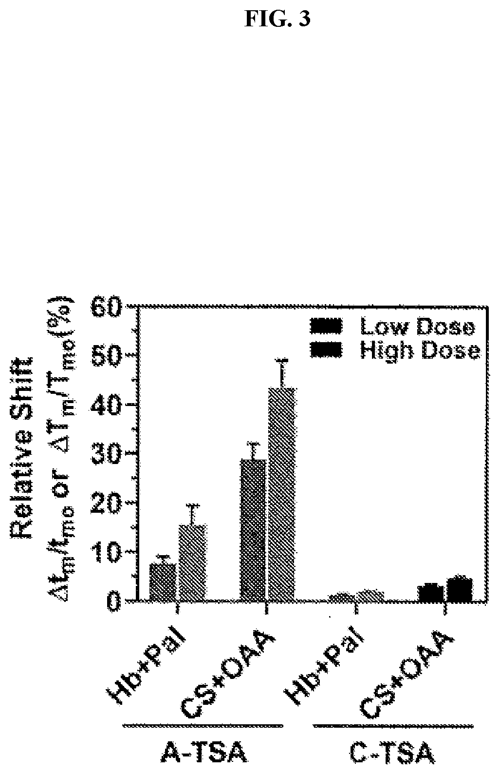
Thermal shift assays (TSAs) have been extensively used to study thermodynamics of proteins and provide an efficient means to assess protein-ligand binding or protein-protein interaction. However, existing TSAs have limitations such as time consuming, labor intensive, or low sensitivity. Here, an acousto-thermal shift assay (A-TSA) is disclosed and is believed to be the first ultrasound enabled TSA which can provide a real-time analysis of protein thermodynamic stability. A-TSA couples unique acoustic mechanisms to achieve protein unfolding, concentration, and measurement on a single microfluidic chip within minutes. Compared to conventional TSA methods, A-TSA provides an ultra-fast (at least 30 times faster), highly sensitive (7-34 folds higher), and label -free monitoring of protein-ligand interactions and protein stability finding applications for protein analysis in biology, medicine and fast diagnosis.

Description

FIELD OF THE INVENTION[0001]The present invention is related to the field of analytical chemistry. In particular, the invention defines a new and improved device and method to detect precise differences in protein secondary, tertiary or quaternary structures. By applying standing acoustic waves to a proteinaceous fluid, differences in protein unfolding characteristics (e.g., melting temperatures) are detected which are beyond the capabilities of conventional thermal shift assays. Such improvements are based upon concomitant protein aggregation that increases local protein concentrations permitting increased temperature shift detection sensitivity.BACKGROUND[0002]Proteins are essential components of organisms and participate in most biological processes. Studying protein dynamics and its interaction with other molecules impact almost every field from fundamental biology to clinical applications.[0003]However, conventional measurement assays and sample preparation takes from many hour...

Claims


Application Information

Patent Timeline
IPC IPC(8): G01N33/68B01L3/00
CPCG01N33/6893B01L3/502761B01L2400/0436B01L2400/0439B01L2300/0645G01N2800/22G01N2001/4094G01N1/4077B01L3/50273B01L7/52B01L2200/0668
Inventor DING, XIAOYUN
Owner THE REGENTS OF THE UNIV OF COLORADO - A BODY CORP
Learn More