Method and apparatus for non-invasive detection of angiogenic and anti-angiogenic activity in living tissue
a living tissue and angiogenic activity technology, applied in the direction of color/spectral properties measurement, diagnostics using light, sensors, etc., can solve the problems of inability to perform a single measurement, prohibitively expensive, and too time-consuming for screening purposes,
- Summary
- Abstract
- Description
- Claims
- Application Information
AI Technical Summary
Problems solved by technology
Method used
Image
Examples
example 2
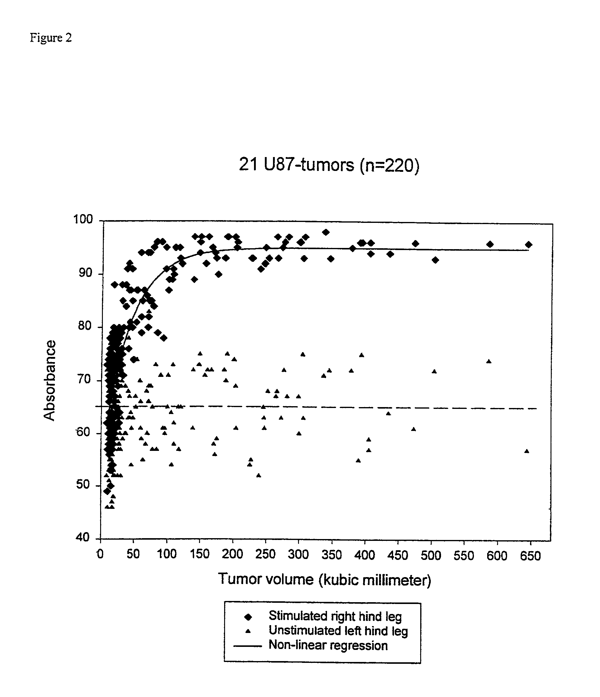
[0051] The apparatus as shown on FIG. 9 was tested in a mouse model with a controlled angiogenic response induced by local application of xenotransplanted human cancer tissue as the source of angiogenic stimulus. Implants with human tissue with very high expression of angiogenic factors (U-87, a human glioblastoma multiform) was repeatedly examined during growth. When the tumor transplants reached a certain size, the absorption reached a maximum (FIG. 2), which appeared to be specific for individual tumor types, and correlated closely to the angiogenic activity as evaluated by expression (protein and MRNA levels) of angiogenic factors and by histological estimates of vessel density. Other tumor lines (U118, U373, 54A, 54B and DMS79), some with a much lower angiogenic activity than U-87, were also tested in a similar experiment. They all reached a specific maximum characteristic of each tumor.
example 3
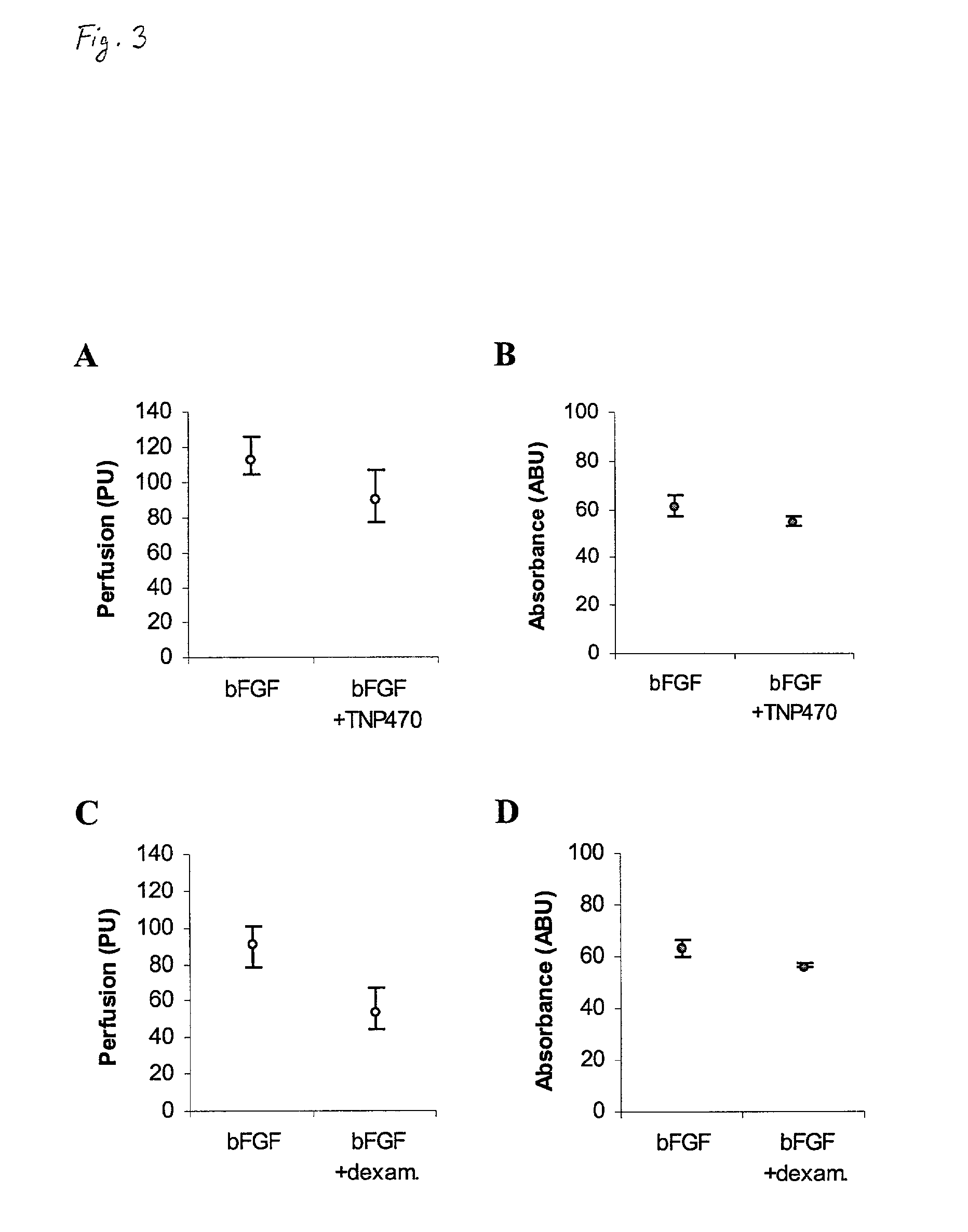
[0052] The ability to detect anti-angiogenic properties of therapeutic agents in these preparations was tested in the same technical settings by comparing treated versus untreated animals. Significant anti-angiogenic activity was demonstrated during treatment with a specific anti-angiogenic compound, TNP-470 (FIGS. 3A and 3B) which is a selective inhibitor of endothelial cell proliferation, and with dexamethasone (FIGS. 3C and 3D) which is a synthetic glucocorticoid analogue.
[0053] FIGS. 3A and 3B show the anti-angiogenic activity after treatment with TNP-470 (7 mg / kg q.d.x7, n=13). LDF (p<0.05) and NIRS (p<0.05) recordings were significantly lower compared to untreated bFGF stimulated animals (n=14). FIGS. 3C and 3D show the anti-angiogenic activity after treatment with dexamethasone (10 mg / kg q.d.x7, n=12), LDF (p<0.001) and NIRS (p<0.001) recordings were significantly lower compared to untreated bFGF stimulated animals (n=14). LDF recordings were performed on day 6, whereas NIRS ...
example 4
[0054] The relationship between non-invasive measures of tumor hemoglobin concentration by NIRS and estimates of vessel density by Chalkley counts was examined in seven tumor lines (gliomas: U87, U118, U373; Small Cell Lung Cancer (SCLC): 54A, 54B, DMS79; rat prostate cancer: MLL) on the hind leg of nude mice. Chalkley counts were obtained from CD31 immunostained cryosections. The NIRS absorbance increased with the tumor size until a tumor line specific plateau was reached from tumor sizes of 100-150 mm.sup.3. FIG. 4 shows that NIRS absorbance and vessel density of the tumors were strongly correlated (r.sub.S=0.77, p<0.001, n=69), thus indicating that NIRS provides a noninvasive estimate of the tumor vessel density that can be used as a reliable estimate of tumor vascularization.
[0055] We also evaluated the correlation between tumor size and NIRS recordings during untreated growth for all seven tumor lines, and during continuous anti-angiogenic treatment with TNP-470 for two tumor l...
PUM
 Abstract
Description
Claims
Application Information
 - R&D
- Intellectual Property
- Life Sciences
- Materials
- Tech Scout
- Unparalleled Data Quality
- Higher Quality Content
- 60% Fewer Hallucinations
Browse by: Latest US Patents, China's latest patents, Technical Efficacy Thesaurus, Application Domain, Technology Topic, Popular Technical Reports.
© 2025 PatSnap. All rights reserved.Legal|Privacy policy|Modern Slavery Act Transparency Statement|Sitemap|About US| Contact US: help@patsnap.com


