Variable angular sampling rate for rotating slat-hole detectors of gamma cameras

Inactive Publication Date: 2002-09-19
MARCONI MEDICAL SYST +1
View PDF0 Cites 5 Cited by
  • Summary
  • Abstract
  • Description
  • Claims
  • Application Information

AI Technical Summary

Benefits of technology

0010] One advantage of the present invention is that it presents a small, relatively light nuclear detector array.
0011] Another advantage is that it presents a solid state nuclear detector array.
0012] Another advantage is that provides un

Problems solved by technology

Unfortunately, there are numerous non-uniformities and inaccuracies when using a large scintillator crystal and an array of photom

Method used


Image

  • Variable angular sampling rate for rotating slat-hole detectors of gamma cameras
  • Variable angular sampling rate for rotating slat-hole detectors of gamma cameras
  • Variable angular sampling rate for rotating slat-hole detectors of gamma cameras

Examples


Embodiment Construction

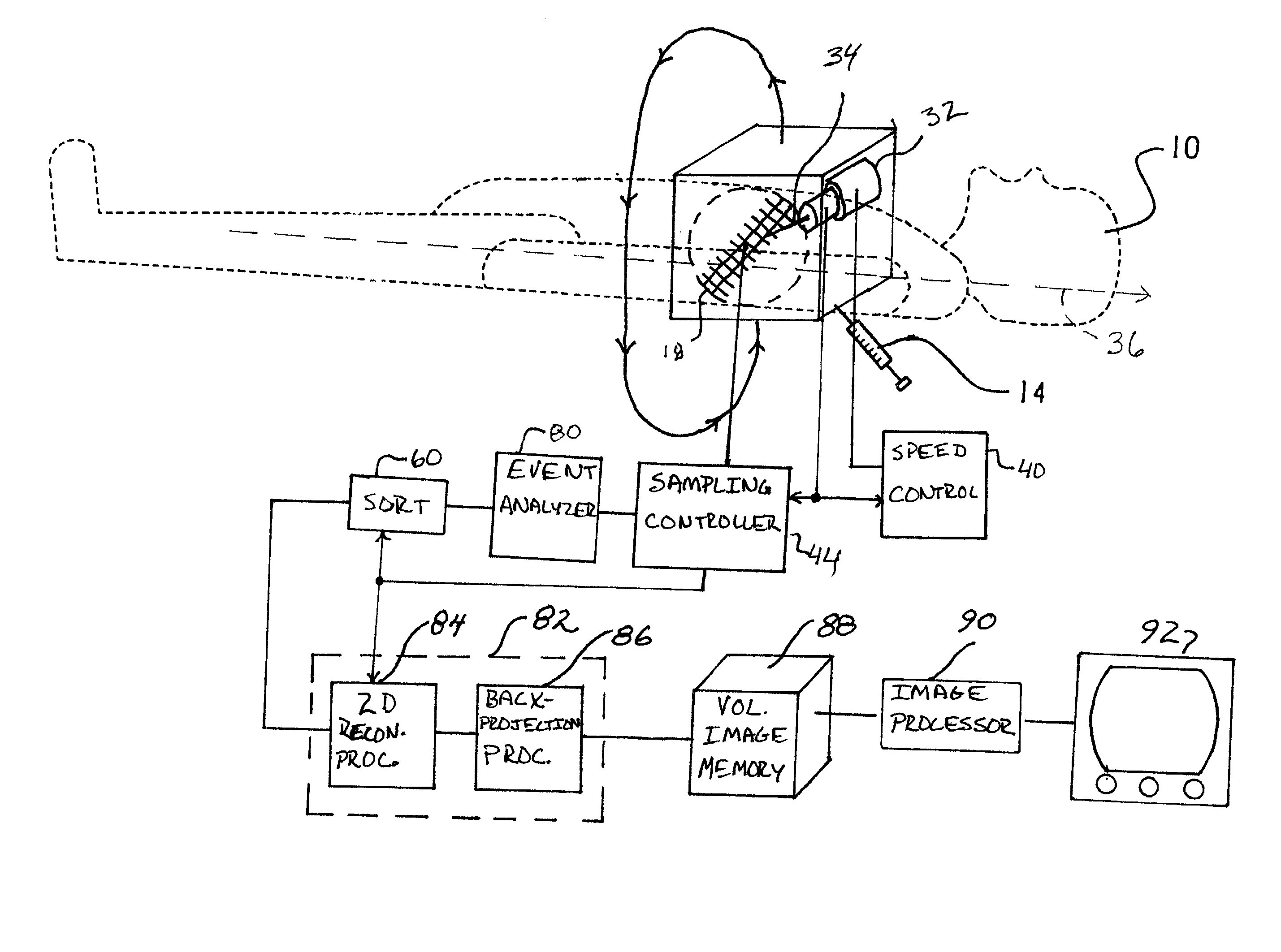
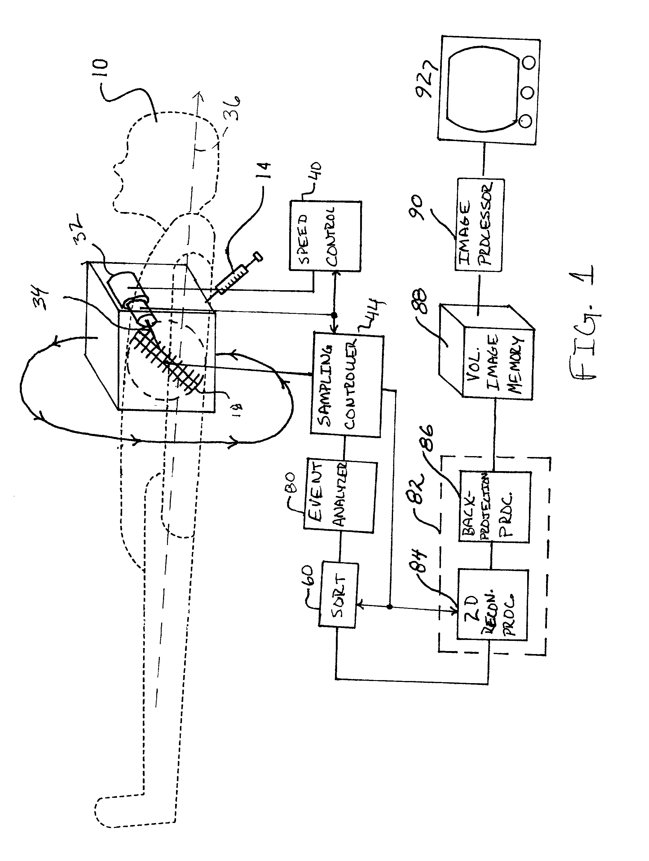
[0023] With reference to FIG. 1, a region of interest of a subject 10 is disposed in an imaging region. In the preferred embodiment, a radiopharmaceutical 14 is injected into the subject, near the region to be imaged. For example, if a physician wanted to view a blockage in the aorta, the isotope would be injected into the bloodstream upstream from the blockage. As another example, the radiopharmaceutical 14 is injected into the circulatory system and its selective absorption by tissue of interest is monitored.

[0024] As quantum physics predicts, atomic nuclei of the radioactive isotope decay over time. Energy is released at the time of decay in the form of a radiation photon, more specifically, a .gamma.-ray of characteristic energy.

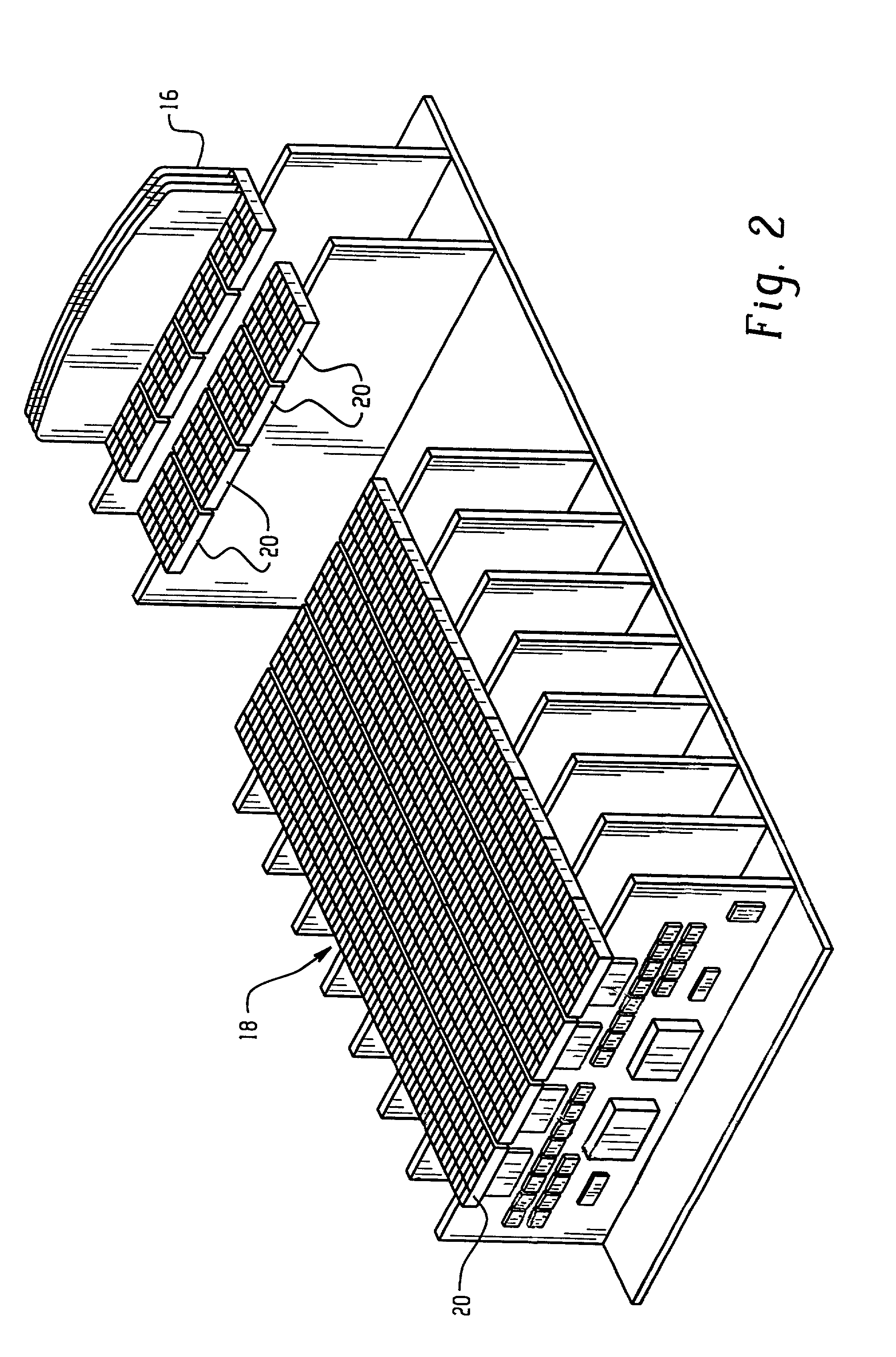
[0025] With reference to FIG. 2, and further reference to FIG. 1, many of the .gamma.-rays produced during an imaging process are lost, propagating in useless directions. However, some of the .gamma.-rays pass through collimators 16, thin tungsten, lead,...


PUM


Abstract

A subject (10) is disposed adjacent a linear detector array (18) of a nuclear camera. The subject (10) is injected with a radioactive isotope (14) and .gamma.-ray emissions indicative of nuclear decay are detected at the detector array (18) as the detector array rotates about an axis of rotation to collect data over a circular field of view. Detectors farther from the axis of rotation are sampled at a higher sampling rate such that the detectors are sampled after a generally constant arc of rotation to correct for angular aliasing. The detector array (18) rotates about the axis of rotation in a 1/sin .theta. pattern with angular offset of the detector array from a longitudinal axis of the subject. This corrects for otherwise uneven sampling. A reconstruction processor (46) reconstructs the identifications of the .gamma.-ray receiving detectors, or other indicators of event detection location, and the digital peak values to generate a spherical image representation.

Description

BACKGROUND OF THE INVENTION[0001] The present invention relates to the diagnostic imaging arts. It finds particular application in conjunction with SPECT nuclear imaging systems and will be described with particular reference thereto. It will be appreciated, however, that the present invention is useful in conjunction with other systems that utilize penetrating radiation, and is not limited to the aforementioned application.[0002] Typically in nuclear imaging, a source of radioactivity is used to provide non-invasive diagnostic images. The source is typically injected into a patient, although external sources are also utilized. Radiation from the source traverses at least a portion of the patient and is detected by radiation detectors.[0003] Typically, a nuclear camera has one, two, or three detector heads. Each head has a large scintillator sheet, such as doped sodium iodide, which converts incident radiation photons into scintillations, i.e. flashes of light. An array of photomult...

Claims


Application Information

Patent Timeline
IPC IPC(8): G01T1/161G01T1/164G01T1/24
CPCG01T1/1644A61B6/037
Inventor ZERG, GENGSHENG LAWRENCEGAGNON, DANIEL
Owner MARCONI MEDICAL SYST
Why Eureka
Try Eureka