Biosensor for use in toxicity assessment and pharmacological screening

a biosensor and toxicity assessment technology, applied in the field of biological sensors, can solve the problems of difficult to relate specific signals to specific functions, inability to control the connection between cells, and inability to reproduce, so as to prevent or inhibit the passage of electrons, the effect of high impedance seal

Inactive Publication Date: 2003-03-20
SCI APPL INT CORP
View PDF1 Cites 20 Cited by
  • Summary
  • Abstract
  • Description
  • Claims
  • Application Information

AI Technical Summary

Benefits of technology

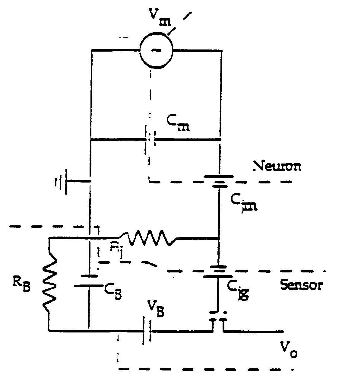
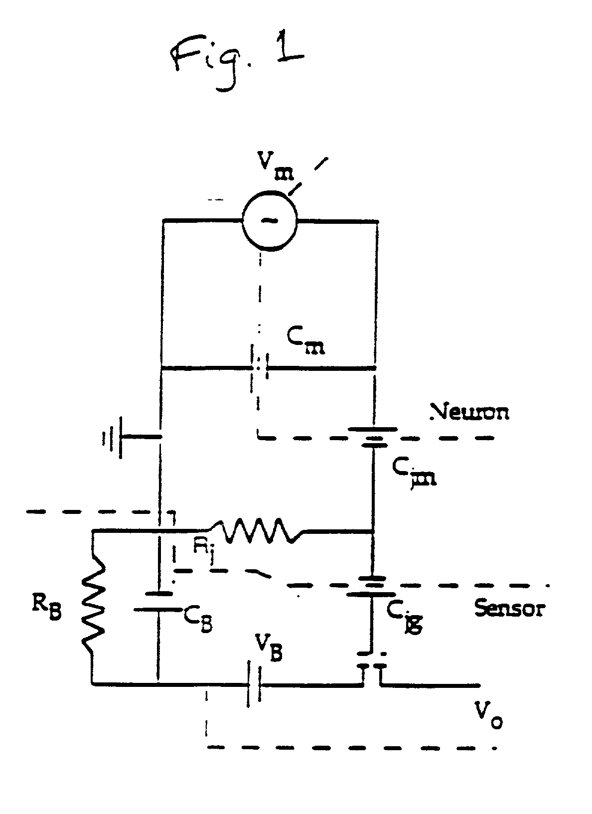
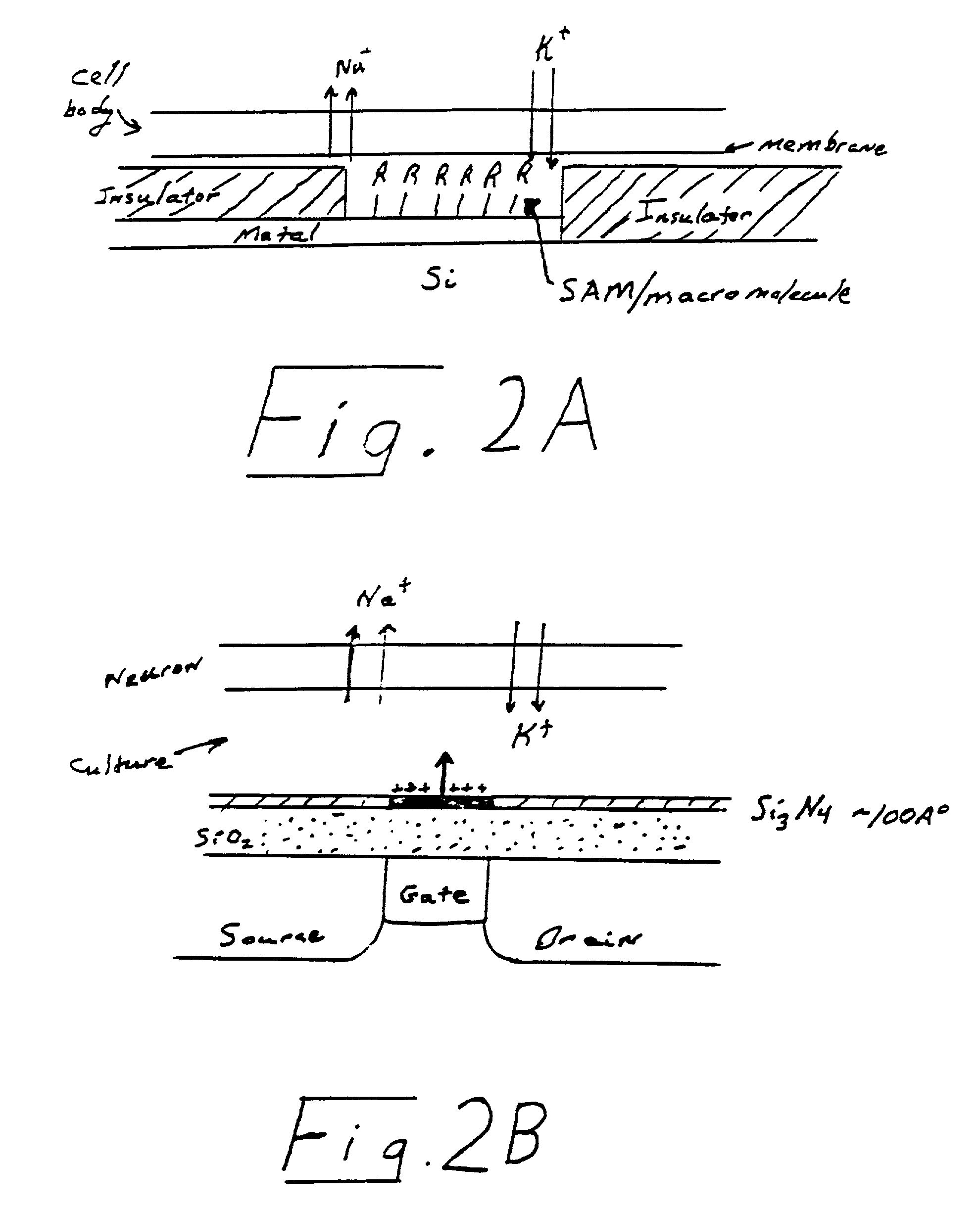
0044] In a preferred embodiment, a biosensor of the invention further comprises at least one insulating or barrier layer interposed between the above-mentioned transducer(s) and the culture medium, which prevents direct contact between the culture medium and the transducer(s). An insulating layer prevents or impedes the passage of electrons between the cell / culture and the transducer, for example, silica and SiC. A barrier layer, as referred to herein, is an insulating layer that also prevents or impedes the passage of ions between the cell / culture and the transducer, for example, Si.sub.4N.sub.3. The axonal or dendritic polarity is provided by the techniques referred to herein, such as through the use of self-assembling monolayers.
0045] Another embodiment of the present biosensor has no insulating layer and a bare metal electrode is in contact with the cell or close to the cell or its processes. The metal is coated with a SAM or biological macromolecule to create a high impedance seal.

Problems solved by technology

However, in the studies to date, neurons in culture are disorganized in the sense that the connections between cells are not controllable and therefore not reproducible.
Historically, this has made it difficult to relate specific signals to specific functions as well as provide for the reproducible system of neuron interconnection necessary to measure small changes in neuron activity associated with changes in the environmental condition of the cells.
However, sensors that can detect unknown agents or agents of intermediate toxicity that affect motor function, cognitive function, and / or other higher order processes are primitive or do not exist.
In this case, however, arbitrary control of axonal / dendritic polarity within topological barriers has not been demonstrated.
Prior methods have not allowed one to hypothesize that a microlithographic approach might be taken to geometrically predispose neurons to send one or more process in a preferred direction.
These approaches have failed to demonstrate the production of isolated features having dimensions in the 1 to 5 micron range, much less establish control over axonal / dendritic cell polarity.
Thus, there has been no report of an effective solution to the problem of controlling the orientation of axonal growth on synthetic surfaces in vitro.
However, no control of neuronal polarity necessary for a reproducible biosensor was obtained in that system.
Such a system requires the placement of a neuron directly on the gate by micromanipulation, thereby being time consuming and open to reproducibility problems.
However, this work does not address the problem of nonspecific absorption of proteins.
However, groups of cells, not individual cells, are "immobilized" by gravitational sedimentation into micromachined silicon wells.
However, there is no suggestion of patterned substrates for the selective adhesion and outgrowth of cells.
These cells, however, did not (and could not be made to) send axons in a preferred or predetermined direction.
Phase contrast micrographs of this mask and immunocytochemical staining, using an axon-specific marker, do not demonstrate the successful induction of axonal / dendritic polarity.
Hence, any features disclosed in these prior publications are ineffective in controlling the direction of axonal outgrowth.
In summary, previous work fails to teach or suggest a biosensor based on rudimentary cognitive function in which changes in the axonal wave potential are monitored and correlated to the presence or absence of a known or unknown bioeffective environmental condition.

Method used


Image

  • Biosensor for use in toxicity assessment and pharmacological screening
  • Biosensor for use in toxicity assessment and pharmacological screening
  • Biosensor for use in toxicity assessment and pharmacological screening

Examples


Embodiment Construction

[0058] According to the principles of the present invention, simple and complex circuits are constructed from mammalian cells, particularly mammalian neuronal cells, oriented in a predetermined way with respect to one another and with respect to one or more transducers to create a biosensor. This sensor can act not only as a screen for known compounds but unknowns as well. These function-based sensors can detect toxins or environmental effects ranging from the obvious (cell death) to those that are more subtle (impairment of function). For instance, substances inhibiting, enhancing, or otherwise affecting the electrical signal output from a natural neuronal network can be found by a screening method utilizing a neuronal network or a biosensor incorporating the network. It is possible under the invention to measure a wide range of responses because neurons and the networks they form are exceedingly sensitive to certain changes in their environment. The development of a solid state de...


PUM

PropertyMeasurementUnit
widthaaaaaaaaaa
widthaaaaaaaaaa
mean diameteraaaaaaaaaa

Abstract

A biosensor for detecting a bioeffecting substance in a test sample includes a cell network of at least one electrically excitable cell provided on a substrate in a predetermined geometry with a predefined axonal/dendritic polarity. The at least one cell is capable of producing a signal in response to the presence of the bioeffecting substance. At least one signal transducer is provided with the substrate in a predetermined geometry and is capable of detecting the signal produced by the cell. A culture medium capable of supporting metabolism of the cell is also provided.

Description

1. FIELD OF THE INVENTION[0002] The present invention relates to a biological sensor useful in the detection of bioeffecting environmental conditions. The biosensor can be used to test for the presence of toxic substances and for screening drugs for pharmacological benefit.2. BACKGROUND OF THE INVENTION[0003] Programs that monitor environmentally hazardous toxins would benefit from sensors based on cell-cell communication, which is one of the lowest levels of cognitive function. One of the reasons they have not been developed to date is the wide range of expertise needed for their reduction to practice. An example of such an application is to assess the effect of waste sources and waste products that may be unknown, especially minor components, which have not been fully identified and characterized. A sensor based on cell-cell communication could screen a processed waste stream to detect toxins or material without requiring that a specific agent be known. Such a biosensor would ensu...

Claims


Application Information

Patent Timeline
Patent Type & Authority Applications(United States)
IPC IPC(8): C12N5/08G01N33/50
CPCB82Y15/00B82Y30/00G01N33/5008G01N33/502G01N33/5058
Inventor HICKMAN, JAMES J.KIRKPATRICK, DOUGLAS A.STENGER, DAVID A.
Owner SCI APPL INT CORP
Why Eureka
Try Eureka