Low back pain and radiculopathy as a result of herniated intervertebral disc represents a major health problem in the United States and all over the world.
A laminectomy is a somewhat destructive procedure, which might cause extensive scarring and long (up to 9 days) hospitalization, with an up to 3-month
postoperative recovery period.
This is
less invasive compared to the laminectomy, though the existing microdiscectomy may still cause some complications similar to those associated with laminectomy, for example,
possible injury to the
nerve root and dural sac, postoperative scarring and relatively long
recovery time.
The method of U.S. Pat. No. 4,545,374 has the following drawbacks: 1) the material is removed from the center of the disc only, thus preventing a surgeon from excising the fragments from the actual herniation site, which may or may not cause recurrent symptoms; 2) this technique is unsuitable for noncontained (or sequestered) herniations, since it does not give a surgeon access to the
epidural space.
Like in the
percutaneous approach described above, one of the disadvantages inherent in this procedure is that it deals with the disc
nucleus, rather than with the herniation itself.
Lasers are both expensive and somewhat tedious to use in these procedures.
Both methods described in U.S. Pat. No. 6,264,650 and in U.S. Pat. No. 6,254,553 make it complicated for a surgeon to focus treatment on the symptomatic site, without affecting the surrounding tissues.
Single-portal MISS methods are limited to the use of one channel at a time.
However, this biportal procedure assumes the second portal to be created from the opposite side to the first portal (bilateral), hence increasing the
operating time, post-operative morbidity, surgeon
exposure to
radiation.
It may also cause excessive removal of disc nuclear tissue, therefore increasing the possible post-operative
stenosis (narrowing) of the
foramen.
These approaches do not provide a comprehensive solution for percutaneous disc surgery.
Oval cannulae, though providing additional space for instruments, are still too restrictive, while more invasive than circular ones.
The method described in U.S. Pat. No. 5,730,754 still needs accurate targeting and it is not sufficiently universal.
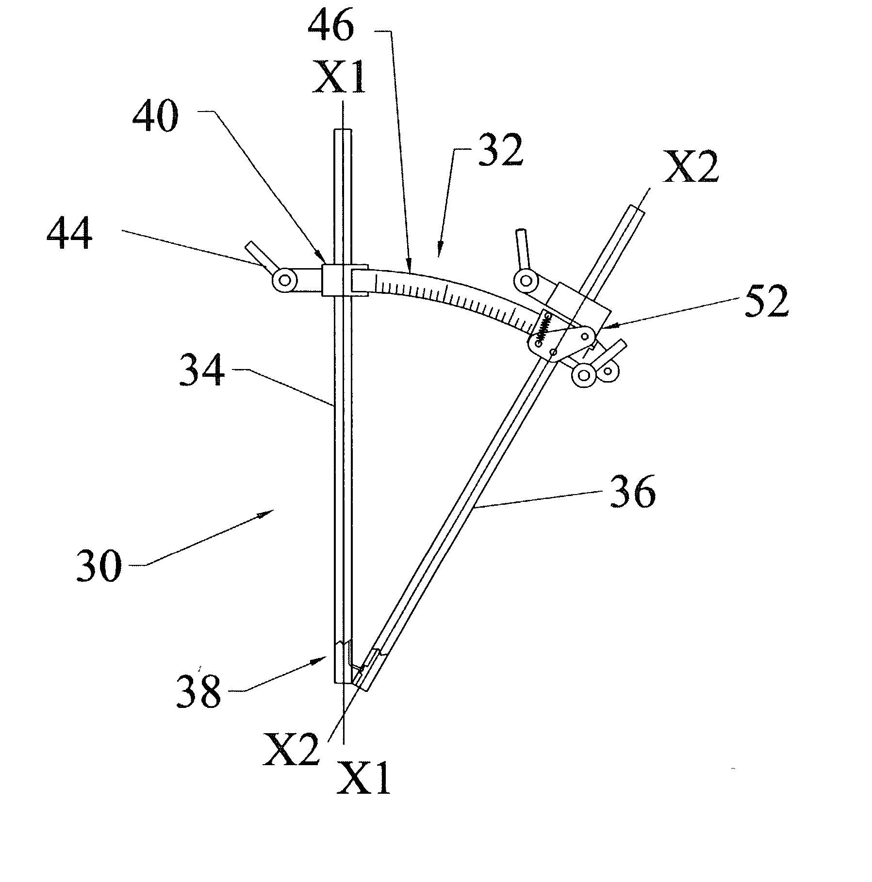
U.S. Pat. No. 5,762,629 allows inserting a second cannula using a special targeting device, but has following disadvantages: 1) the targeting device is rigid and does not allow the flexibility required by a surgeon for the formation of an angle between inserted cannulae; 2) after cannulae are inserted, the targeting device is removed, leaving the cannulae completely unlinked, so that a surgeon cannot keep them interrelated.
In case the position of one of the inserted cannulae should be temporarily changed, it becomes a problem to reorient them, especially when more than two cannulae are used for the surgery.
Another common
disadvantage of the existing devices for the
percutaneous surgery is that they require the operation to be carried out under X-
ray monitoring at all steps of the surgery, i.e., during
insertion of each additional cannula and occasionally during the procedure itself.

