
Novel chimeric proteins and methods for using the same
a technology of chimeric proteins and chimeric proteins, applied in the field of new chimeric proteins and methods for using the same, can solve the problems of naturally processed sfasl molecule form trimers, inflammatory reaction and tissue damage, and autoimmune diseases
- Summary
- Abstract
- Description
- Claims
- Application Information
AI Technical Summary
Problems solved by technology
Method used
Image

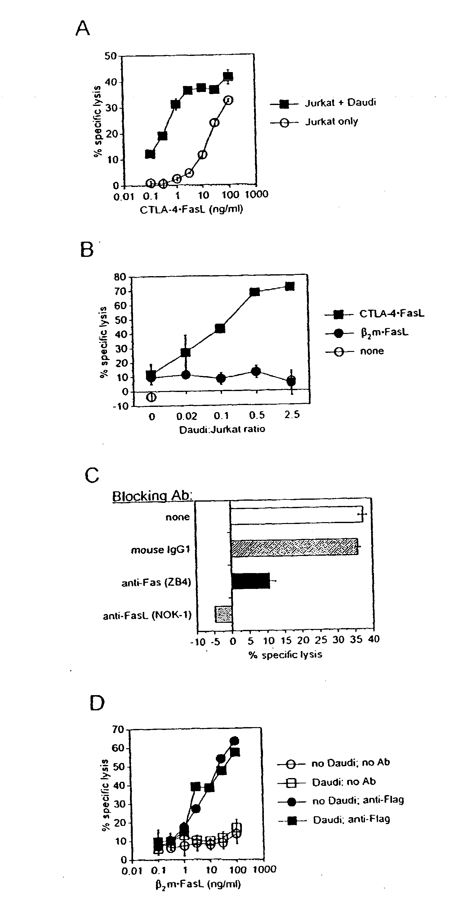
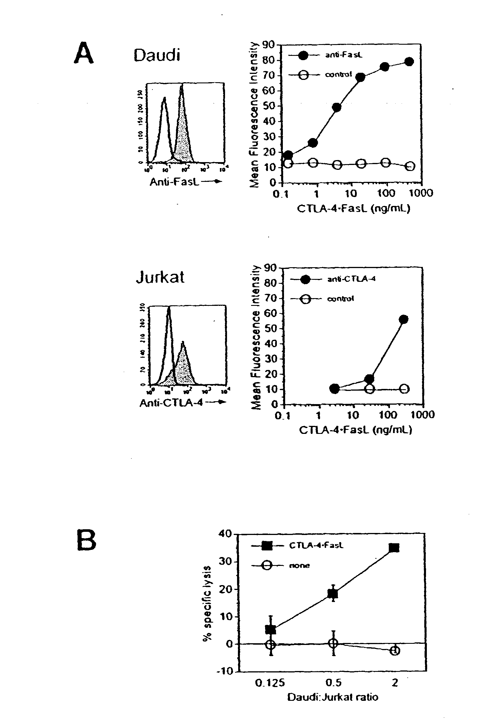
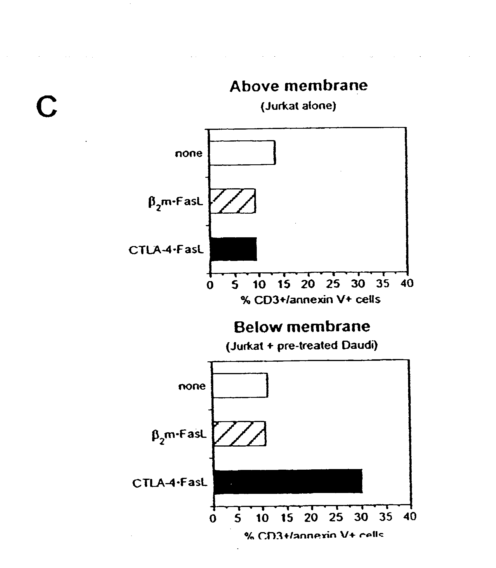
Examples
example 1
[0054] Plasmid Construction and Transfection
[0055] Two targeting domains were genetically chimerized with sequences encoding soluble FasL: extracellular domain sequence from human CTLA-4 and human beta-2-microglobulin (B2M). The chimerization of FasL with CTLA-4 within CTLA-4.multidot.FasL allows binding to resident B7-1 / B7-2 (CD80 / CD86) molecules on APC, in addition to the pro-apoptotic activity of FasL. The soluble B2M.multidot.FasL protein was developed as a control. For expression of chimeric sFasL-containing proteins, recombinant expression plasmids were constructed and expressing cell lines selected from transfected 293 human embryonic kidney cells.
[0056] The cDNAs for human FasL have been described previously by Takahashi (1994). Synthetic oligonucleotides were purchased from Genosys, Inc. (The Woodlands, Texas). DNA primers were designed to replace the stop codon of human B2M with a Hind III restriction site. DNA primers for the generation of a human B2M sequence encoding am...
example 2
[0071] Role of cell-to-cell contact in mediating CTLA-4.multidot.FasL trans-effector activity
[0072] Soluble CTLA-4 polypeptide derivatives, such as CTLA-4.multidot.Ig, bind to cells expressing CD80 and / or CD86. To test whether the binding of CTLA-4.multidot.FasL molecules to CD80 / CD86-positive cells potentiate Jurkat cell apoptosis, the ability of CTLA-4.multidot.FasL to bind Daudi and Jurkat cells was assessed. CD80 / CD86-positive Daudi cells were pre-incubated with CTLA-4.multidot.FasL-containing cell supernatants and processed for indirect immunofluorescence and flow cytometry using a FasL-specific monoclonal antibody or an isotype-matched control antibody. More specifically, pre-incubation was performed at 0.degree. C. for 30-45 minutes. After two washes, cells were incubated with anti-FasL antibody (NOK-1, J) or isotype-matched control antibody (Control, E) at 10 mg / ml for 30-45 minutes at 37.degree. C. Following two washes, cells were incubated with FITC-conjugated goat anti-mo...
example 3
[0075] CTLA-4.circle-solid.FasL was tested for the capacity to inhibit the polyclonal proliferation of human peripheral blood T cells to mitogenic anti-CD3 antibody. In 96-well U bottom tissue culture plates, 2.times.10.sup.5 PBMC / well of freshly isolated human peripheral blood mononuclear cells (PBMC) from two individual donors were cultured with 1 ng / ml CD3-specific antibody (OKT3) in the absence or presence of varying amounts of immunoaffinity-purified CTLA-4.circle-solid.FasL. Assays were performed in triplicate. Freshly isolated human peripheral blood mononuclear cells from two individual donors were cultured with 1 ng / ml CD3-specific antibody (OKT3) in the absence or presence of varying amounts of immunoaffinity-purified CTLA-4.circle-solid.FasL. For comparison, 10,000 ng / ml CTLA-4.circle-solid.Ig and recombinant human soluble FasL were also titrated into parallel cultures. After 36 hours and at 24-hour intervals for three additional days, culture wells were pulsed with .sup.3...
PUM
| Property | Measurement | Unit |
|---|---|---|
| molecular weight | aaaaa | aaaaa |
| pore size | aaaaa | aaaaa |
| concentration | aaaaa | aaaaa |
Abstract
Description
Claims
Application Information