Elongated markers for soft tissue volume identification

a soft tissue volume and marker technology, applied in the field of elongated markers for soft tissue volume identification, can solve the problems of not being able to follow the change of shape of said organ, markers are of no use, diagnostic tools cannot distinguish between malignant and benign tumors,

Active Publication Date: 2004-04-15
RADIOMED
View PDF24 Cites 91 Cited by
  • Summary
  • Abstract
  • Description
  • Claims
  • Application Information

AI Technical Summary

Benefits of technology

[0014] It is another object of the present invention to provide method for precisely localising an organ or tumor to be treated with a radiation beam, and a marker adapted for use in said method.
[0017] The present invention is related, in one aspect, to an interstitial marker for localizing an organ, tumor or tumor bed within a mammalian body. The marker has a proximal end, a distal end, and a continuous intervening length. At least a portion of the intervening length is visible under at least one imaging modality and has a flexibility such that said marker follows movements and changes of shape of said organ, tumor or tumor bed. In a preferred embodiment, the interstitial marker has a ratio of said length to its diameter of 10 or higher. Depending on the application, said ratio may even be of 250 or higher. The interstitial marker may comprise a flexible wire, and said wire may be in form of a helical coil, whereby an advantageous flexibility is obtained. The helical coil marker may comprise sections of different pitch. Sections of different pitch have different characteristics as to visibility under different imaging modalities, and also have different mechanical characteristics as to longitudinal and lateral flexibility. At least portions of the marker may be made of a radiopaque material. They may be visible under diagnostic x-ray imaging, eg x-rays having an energy in the range of 100 keV. They may also advantageously be visible under higher energy x-rays used in treatment beams, eg x-rays in the range of 3 to 50 MeV. They also preferably may be visible under ultrasound imaging. The marker may include one or more tissue anchors at opposite ends thereof, and / or at intermediate points to fix the marker in the organ, tumor or tumor bed. The marker will thereby follow changes of size and shape, especially longitudinal size changes. Other means for anchoring to said organ, tumor or tumor bed may be used, e.g. a structure of the lateral surface of the marker improving the grip of the marker to the tissue, such as the ridges on the outer surface of an helical coil. The marker is preferably made of a biocompatible material suitable for permanent implantation. Such material may be rhodium, platinum, iridium, tantalum, titanium, silver, gold, nickel, alloys containing these metals and stainless steel.

Problems solved by technology

The diagnostic tool does not allow making a distinction between a malignant and a benign tumor.
However, for soft tissues, which are not in a fixed position with respect to bone structures, these markers are of no avail.
It is however not possible to follow the change of shape of said organ.
There is typically no detailed knowledge of the behavior of the tissue between the markers.
This can lead to misinterpretation of the target volume change or deformation if the tissue or organ is being subjected to either external or internal forces.
However, such seeds have two drawbacks: they can drift away from their intended location, and they give poor visibility to ultrasound imaging.
These devices have, as a result of their size and rigidity, many drawback for use as interstitial markers: (i) they can not be introduced through a minimally invasive intervention, such as a fine needle; (ii) they may not be readily accepted as permanent marker, left in the body, in case the biopsy does not reveal the tumor to be malignant; (iii) they lack the lateral and axial flexibility that is required for permanent insertion in soft tissues that may shrink or swell under the influence of outside factors; (iv) they may mask and conceal features of the lesion that should remain visible to the therapist.
The placement of a set of such clips does not allow the follow-up of the shape of the cavity.
The presence of a filler body may not improve the healing process of the biopsy site.

Method used


Image

  • Elongated markers for soft tissue volume identification
  • Elongated markers for soft tissue volume identification
  • Elongated markers for soft tissue volume identification

Examples


example 2

[0046] Designed primarily for diagnostic x-ray, fluoroscope or ultrasound

2 Material Platinum Helix Outer Diameter 500 .mu.m Helix Inner Diameter 350 .mu.m Circular wire 75 .mu.m diameter Wire Pitch 90 + / - 9 .mu.m

example 3

[0047] Primarily designed for Portal Imaging (high energy x-rays)

3 Material Gold Helix Outer Diameter 2.0 mm Helix Inner Diameter 1.2 mm Wire Diameter 0.4 mm Wire Pitch 0.48 + / - 0.08 mm

[0048] The lateral flexibility obtained by using a coil according to the invention is very high. This flexibility is easily determined by measuring the droop of a length of helical coil fastened horizontaly from one end. The other end droops in response to its own weight. It was determined that a coil made of stainless steel, having an outer diameter of 350 .mu.m, a rectangular cross section wire of 200 .mu.m by 50 .mu.m, a 220 .mu.m pitch, and a free length of 35.5 mm, droops by 2.9 mm.

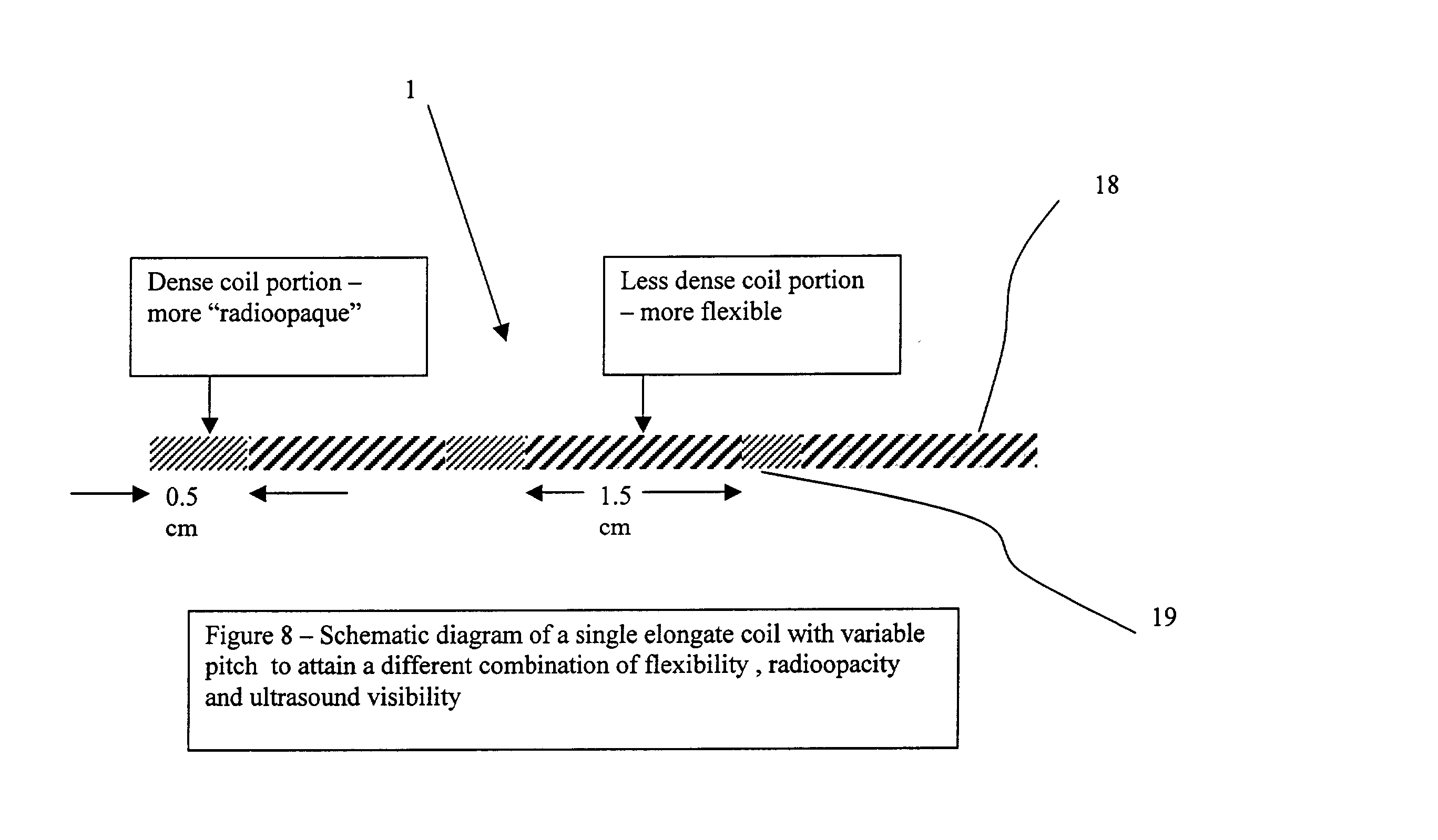
[0049] FIG. 8 represents a preferred embodiment of a coiled marker 1, having sections of different pitch. Sections of high pitch 18 (less dense coils) provide a good flexibility, while sections of low pitch 19 (dense coils) provide good x-ray and ultrasound visibility.

[0050] FIGS. 9a-9d represent images of the marker c...


PUM


Abstract

The present invention is related to an interstitial marker for localizing an organ, tumor or tumor bed within a mammalian body wherein said marker has a proximal end, a distal end, and a continuous intervening length, at least a portion of the intervening length of said marker being visible under at least one imaging modality and having a flexibility such that said marker follows movements and changes of shape of said organ, tumor or tumor bed.

Description

[0001] The present invention is related to a device and method for marking, localizing and tracing the absolute position, motion and / or distortion of organs and regions or voids in organs, particularly in flexible tissue in human and animal subjects. The invention is further related to methods of use of the device of the invention.STATE OF THE ART[0002] Interstitial markers are used in many medical applications for localizing and following-up regions of body.[0003] A first typical application of interstitial markers is in the following context: a tissue abnormality is detected by a known diagnostic tool, such as ultrasonography, PET, SPECT, CT scan, mammogram, radiography or manual palpation. The diagnostic tool does not allow making a distinction between a malignant and a benign tumor. A biopsy is performed in order to analyze the tumor. A marker is then inserted at the location of the biopsy, for later precise treatment of the tumor, if necessary. Breast cancer diagnosis and treat...

Claims


Application Information

Patent Timeline
Patent Type & Authority Applications(United States)
IPC IPC(8): A61B19/00
CPCA61B19/54A61B90/39
Inventor SIOSHANSI, PIRANBRICAULT, RAYMOND J.
Owner RADIOMED
Why Eureka
Try Eureka